7Z1K

Crystal structure of the SPOC domain of human SHARP (SPEN) in complex with RNA polymerase II CTD heptapeptide phosphorylated on Ser5


Experimental Data Snapshot

  • Method: X-RAY DIFFRACTION
  • Resolution: 1.55 Å
  • R-Value Free: 0.247 
  • R-Value Work: 0.215 
  • R-Value Observed: 0.217 

wwPDB Validation   3D Report Full Report


This is version 1.2 of the entry. See complete history


Literature

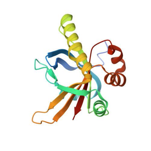
The SPOC domain is a phosphoserine binding module that bridges transcription machinery with co- and post-transcriptional regulators.

Appel, L.M.Franke, V.Benedum, J.Grishkovskaya, I.Strobl, X.Polyansky, A.Ammann, G.Platzer, S.Neudolt, A.Wunder, A.Walch, L.Kaiser, S.Zagrovic, B.Djinovic-Carugo, K.Akalin, A.Slade, D.

(2023) Nat Commun 14: 166-166

  • DOI: https://doi.org/10.1038/s41467-023-35853-1
  • Primary Citation of Related Structures:  
    7Z1K, 7Z27

  • PubMed Abstract: 

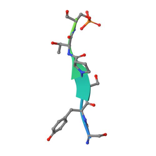
    The heptad repeats of the C-terminal domain (CTD) of RNA polymerase II (Pol II) are extensively modified throughout the transcription cycle. The CTD coordinates RNA synthesis and processing by recruiting transcription regulators as well as RNA capping, splicing and 3'end processing factors. The SPOC domain of PHF3 was recently identified as a CTD reader domain specifically binding to phosphorylated serine-2 residues in adjacent CTD repeats. Here, we establish the SPOC domains of the human proteins DIDO, SHARP (also known as SPEN) and RBM15 as phosphoserine binding modules that can act as CTD readers but also recognize other phosphorylated binding partners. We report the crystal structure of SHARP SPOC in complex with CTD and identify the molecular determinants for its specific binding to phosphorylated serine-5. PHF3 and DIDO SPOC domains preferentially interact with the Pol II elongation complex, while RBM15 and SHARP SPOC domains engage with writers and readers of m 6 A, the most abundant RNA modification. RBM15 positively regulates m 6 A levels and mRNA stability in a SPOC-dependent manner, while SHARP SPOC is essential for its localization to inactive X-chromosomes. Our findings suggest that the SPOC domain is a major interface between the transcription machinery and regulators of transcription and co-transcriptional processes.


  • Organizational Affiliation

    Department of Radiation Oncology, Medical University of Vienna, Währinger Gürtel 18-20, 1090, Vienna, Austria.


Macromolecules
Find similar proteins by:  (by identity cutoff)  |  3D Structure
Entity ID: 1
MoleculeChains Sequence LengthOrganismDetailsImage
Msx2-interacting protein172Homo sapiensMutation(s): 0 
Gene Names: SPENKIAA0929MINTSHARP
UniProt & NIH Common Fund Data Resources
Find proteins for Q96T58 (Homo sapiens)
Explore Q96T58 
Go to UniProtKB:  Q96T58
PHAROS:  Q96T58
GTEx:  ENSG00000065526 
Entity Groups  
Sequence Clusters30% Identity50% Identity70% Identity90% Identity95% Identity100% Identity
UniProt GroupQ96T58
Sequence Annotations
Expand
  • Reference Sequence

Find similar proteins by:  Sequence   |   3D Structure  

Entity ID: 2
MoleculeChains Sequence LengthOrganismDetailsImage
SER-TYR-SER-PRO-THR-SEP16Homo sapiensMutation(s): 0 
UniProt & NIH Common Fund Data Resources
Find proteins for P24928 (Homo sapiens)
Explore P24928 
Go to UniProtKB:  P24928
PHAROS:  P24928
Entity Groups  
Sequence Clusters30% Identity50% Identity70% Identity90% Identity95% Identity100% Identity
UniProt GroupP24928
Sequence Annotations
Expand
  • Reference Sequence
Small Molecules
Modified Residues  1 Unique
IDChains TypeFormula2D DiagramParent
SEP
Query on SEP
B
L-PEPTIDE LINKINGC3 H8 N O6 PSER
Experimental Data & Validation

Experimental Data

  • Method: X-RAY DIFFRACTION
  • Resolution: 1.55 Å
  • R-Value Free: 0.247 
  • R-Value Work: 0.215 
  • R-Value Observed: 0.217 
  • Space Group: P 21 21 21
Unit Cell:
Length ( Å )Angle ( ˚ )
a = 43.327α = 90
b = 60.517β = 90
c = 68.273γ = 90
Software Package:
Software NamePurpose
PHENIXrefinement
PHENIXrefinement
XDSdata reduction
Aimlessdata scaling
PHASERphasing

Structure Validation

View Full Validation Report


Entry History & Funding Information

Deposition Data


Funding OrganizationLocationGrant Number
Austrian Science FundAustria--

Revision History  (Full details and data files)

  • Version 1.0: 2022-12-07
    Type: Initial release
  • Version 1.1: 2023-01-25
    Changes: Database references
  • Version 1.2: 2024-02-07
    Changes: Data collection, Refinement description