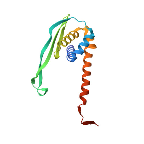
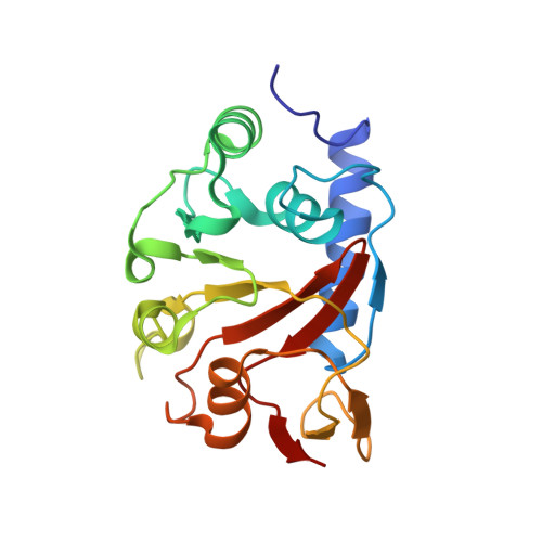
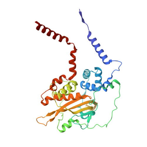
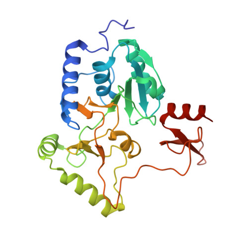
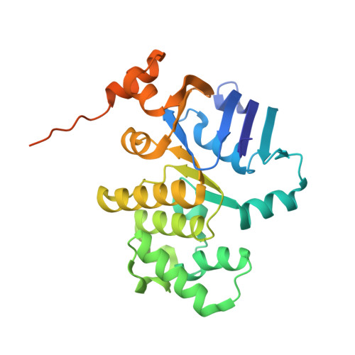
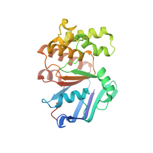
7Z15

E. coli C-P lyase bound to a PhnK/PhnL dual ABC dimer and ADP + Pi


Experimental Data Snapshot

  • Method: ELECTRON MICROSCOPY
  • Resolution: 1.93 Å
  • Aggregation State: PARTICLE 
  • Reconstruction Method: SINGLE PARTICLE 

wwPDB Validation   3D Report Full Report


This is version 1.1 of the entry. See complete history


Literature

Structural remodelling of the carbon-phosphorus lyase machinery by a dual ABC ATPase.

Amstrup, S.K.Ong, S.C.Sofos, N.Karlsen, J.L.Skjerning, R.B.Boesen, T.Enghild, J.J.Hove-Jensen, B.Brodersen, D.E.

(2023) Nat Commun 14: 1001-1001

  • DOI: https://doi.org/10.1038/s41467-023-36604-y
  • Primary Citation of Related Structures:  
    7Z15, 7Z16, 7Z17, 7Z18, 7Z19

  • PubMed Abstract: 

    In Escherichia coli, the 14-cistron phn operon encoding carbon-phosphorus lyase allows for utilisation of phosphorus from a wide range of stable phosphonate compounds containing a C-P bond. As part of a complex, multi-step pathway, the PhnJ subunit was shown to cleave the C-P bond via a radical mechanism, however, the details of the reaction could not immediately be reconciled with the crystal structure of a 220 kDa PhnGHIJ C-P lyase core complex, leaving a significant gap in our understanding of phosphonate breakdown in bacteria. Here, we show using single-particle cryogenic electron microscopy that PhnJ mediates binding of a double dimer of the ATP-binding cassette proteins, PhnK and PhnL, to the core complex. ATP hydrolysis induces drastic structural remodelling leading to opening of the core complex and reconfiguration of a metal-binding and putative active site located at the interface between the PhnI and PhnJ subunits.


  • Organizational Affiliation

    Department of Molecular Biology and Genetics, Aarhus University, Universitetsbyen 81, DK-8000, Aarhus C, Denmark.


Macromolecules
Find similar proteins by:  (by identity cutoff)  |  3D Structure
Entity ID: 1
MoleculeChains Sequence LengthOrganismDetailsImage
Alpha-D-ribose 1-methylphosphonate 5-triphosphate synthase subunit PhnG
A, E
150Escherichia coliMutation(s): 0 
Gene Names: phnGb4101JW4062
EC: 2.7.8.37
UniProt
Find proteins for P16685 (Escherichia coli (strain K12))
Explore P16685 
Go to UniProtKB:  P16685
Entity Groups  
Sequence Clusters30% Identity50% Identity70% Identity90% Identity95% Identity100% Identity
UniProt GroupP16685
Sequence Annotations
Expand
  • Reference Sequence
Find similar proteins by:  (by identity cutoff)  |  3D Structure
Entity ID: 2
MoleculeChains Sequence LengthOrganismDetailsImage
Alpha-D-ribose 1-methylphosphonate 5-triphosphate synthase subunit PhnH
B, F
194Escherichia coliMutation(s): 0 
Gene Names: phnHb4100JW4061
EC: 2.7.8.37
UniProt
Find proteins for P16686 (Escherichia coli (strain K12))
Explore P16686 
Go to UniProtKB:  P16686
Entity Groups  
Sequence Clusters30% Identity50% Identity70% Identity90% Identity95% Identity100% Identity
UniProt GroupP16686
Sequence Annotations
Expand
  • Reference Sequence
Find similar proteins by:  (by identity cutoff)  |  3D Structure
Entity ID: 3
MoleculeChains Sequence LengthOrganismDetailsImage
Alpha-D-ribose 1-methylphosphonate 5-triphosphate synthase subunit PhnI
C, G
354Escherichia coliMutation(s): 0 
Gene Names: phnIb4099JW4060
EC: 2.7.8.37
UniProt
Find proteins for P16687 (Escherichia coli (strain K12))
Explore P16687 
Go to UniProtKB:  P16687
Entity Groups  
Sequence Clusters30% Identity50% Identity70% Identity90% Identity95% Identity100% Identity
UniProt GroupP16687
Sequence Annotations
Expand
  • Reference Sequence
Find similar proteins by:  (by identity cutoff)  |  3D Structure
Entity ID: 4
MoleculeChains Sequence LengthOrganismDetailsImage
Alpha-D-ribose 1-methylphosphonate 5-phosphate C-P lyase
D, H
281Escherichia coliMutation(s): 0 
Gene Names: phnJb4098JW4059
EC: 4.7.1.1
UniProt
Find proteins for P16688 (Escherichia coli (strain K12))
Explore P16688 
Go to UniProtKB:  P16688
Entity Groups  
Sequence Clusters30% Identity50% Identity70% Identity90% Identity95% Identity100% Identity
UniProt GroupP16688
Sequence Annotations
Expand
  • Reference Sequence
Find similar proteins by:  (by identity cutoff)  |  3D Structure
Entity ID: 5
MoleculeChains Sequence LengthOrganismDetailsImage
Putative phosphonates utilization ATP-binding protein PhnK
I, J
291Escherichia coliMutation(s): 0 
Gene Names: phnKb4097JW5727
UniProt
Find proteins for P16678 (Escherichia coli (strain K12))
Explore P16678 
Go to UniProtKB:  P16678
Entity Groups  
Sequence Clusters30% Identity50% Identity70% Identity90% Identity95% Identity100% Identity
UniProt GroupP16678
Sequence Annotations
Expand
  • Reference Sequence
Find similar proteins by:  (by identity cutoff)  |  3D Structure
Entity ID: 6
MoleculeChains Sequence LengthOrganismDetailsImage
Alpha-D-ribose 1-methylphosphonate 5-triphosphate synthase subunit PhnL
K, L
226Escherichia coliMutation(s): 0 
Gene Names: phnLb4096JW4057
EC: 2.7.8.37
UniProt
Find proteins for P16679 (Escherichia coli (strain K12))
Explore P16679 
Go to UniProtKB:  P16679
Entity Groups  
Sequence Clusters30% Identity50% Identity70% Identity90% Identity95% Identity100% Identity
UniProt GroupP16679
Sequence Annotations
Expand
  • Reference Sequence
Small Molecules
Ligands 6 Unique
IDChains Name / Formula / InChI Key2D Diagram3D Interactions
ATP
Query on ATP

Download Ideal Coordinates CCD File 
AA [auth L],
Y [auth K]
ADENOSINE-5'-TRIPHOSPHATE
C10 H16 N5 O13 P3
ZKHQWZAMYRWXGA-KQYNXXCUSA-N
ADP (Subject of Investigation/LOI)
Query on ADP

Download Ideal Coordinates CCD File 
S [auth I],
V [auth J]
ADENOSINE-5'-DIPHOSPHATE
C10 H15 N5 O10 P2
XTWYTFMLZFPYCI-KQYNXXCUSA-N
I9X (Subject of Investigation/LOI)
Query on I9X

Download Ideal Coordinates CCD File 
O [auth D],
R [auth H]
alpha-D-ribose-1,2-cyclic-phosphate-5-phosphate
C5 H10 O10 P2
OXGUIUWFXGIWNM-TXICZTDVSA-N
PO4 (Subject of Investigation/LOI)
Query on PO4

Download Ideal Coordinates CCD File 
T [auth I],
W [auth J]
PHOSPHATE ION
O4 P
NBIIXXVUZAFLBC-UHFFFAOYSA-K
ZN (Subject of Investigation/LOI)
Query on ZN

Download Ideal Coordinates CCD File 
M [auth C],
N [auth D],
P [auth G],
Q [auth H]
ZINC ION
Zn
PTFCDOFLOPIGGS-UHFFFAOYSA-N
MG
Query on MG

Download Ideal Coordinates CCD File 
BA [auth L],
U [auth I],
X [auth J],
Z [auth K]
MAGNESIUM ION
Mg
JLVVSXFLKOJNIY-UHFFFAOYSA-N
Experimental Data & Validation

Experimental Data

  • Method: ELECTRON MICROSCOPY
  • Resolution: 1.93 Å
  • Aggregation State: PARTICLE 
  • Reconstruction Method: SINGLE PARTICLE 
EM Software:
TaskSoftware PackageVersion
RECONSTRUCTIONcryoSPARC3.2
MODEL REFINEMENTPHENIX

Structure Validation

View Full Validation Report


Entry History & Funding Information

Deposition Data


Funding OrganizationLocationGrant Number
Novo Nordisk FoundationDenmarkNNF18OC0030646

Revision History  (Full details and data files)

  • Version 1.0: 2022-06-22
    Type: Initial release
  • Version 1.1: 2023-03-08
    Changes: Database references, Refinement description