7YLG

Crystal structure of the chicken Toll-like receptor 15 TIR domain (glutathione adduct)


Experimental Data Snapshot

  • Method: X-RAY DIFFRACTION
  • Resolution: 1.80 Å
  • R-Value Free: 0.242 
  • R-Value Work: 0.214 
  • R-Value Observed: 0.215 

wwPDB Validation   3D Report Full Report


Ligand Structure Quality Assessment 


This is version 1.1 of the entry. See complete history


Literature

Structural analysis of the Toll-like receptor 15 TIR domain.

Ko, K.Y.Song, W.S.Park, J.Lee, G.S.Yoon, S.I.

(2023) IUCrJ 10: 352-362

  • DOI: https://doi.org/10.1107/S2052252523002956
  • Primary Citation of Related Structures:  
    7YLF, 7YLG

  • PubMed Abstract: 

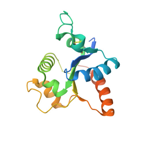
    Toll-like receptors (TLRs) activate innate immunity in response to pathogen-associated molecular patterns (PAMPs). The ectodomain of a TLR directly senses a PAMP and the intracellular TIR domain dimerizes to initiate a signaling cascade. The TIR domains of TLR6 and TLR10, which belong to the TLR1 subfamily, have been structurally characterized in a dimer, whereas those of other subfamilies, including TLR15, have not been explored at the structural or molecular level. TLR15 is a TLR unique to birds and reptiles that responds to virulence-associated fungal and bacterial proteases. To reveal how the TLR15 TIR domain (TLR15 TIR ) triggers signaling, the crystal structure of TLR15 TIR was determined in a dimeric form and a mutational study was performed. TLR15 TIR forms a one-domain structure in which a five-stranded β-sheet is decorated by α-helices, as shown for TLR1 subfamily members. TLR15 TIR exhibits substantial structural differences from other TLRs at the BB and DD loops and αC2 helix that are involved in dimerization. As a result, TLR15 TIR is likely to form a dimeric structure that is unique in its intersubunit orientation and the contribution of each dimerizing region. Further comparative analysis of TIR structures and sequences provides insights into the recruitment of a signaling adaptor protein by TLR15 TIR .


  • Organizational Affiliation

    Division of Biomedical Convergence, Kangwon National University, Chuncheon, 24341, Republic of Korea.


Macromolecules
Find similar proteins by:  (by identity cutoff)  |  3D Structure
Entity ID: 1
MoleculeChains Sequence LengthOrganismDetailsImage
Toll-like receptor 2176Gallus gallusMutation(s): 0 
EC: 3.2.2.6
UniProt
Find proteins for H2D5F4 (Gallus gallus)
Explore H2D5F4 
Go to UniProtKB:  H2D5F4
Entity Groups  
Sequence Clusters30% Identity50% Identity70% Identity90% Identity95% Identity100% Identity
UniProt GroupH2D5F4
Sequence Annotations
Expand
  • Reference Sequence
Small Molecules
Ligands 1 Unique
IDChains Name / Formula / InChI Key2D Diagram3D Interactions
GSH (Subject of Investigation/LOI)
Query on GSH

Download Ideal Coordinates CCD File 
B [auth A]GLUTATHIONE
C10 H17 N3 O6 S
RWSXRVCMGQZWBV-WDSKDSINSA-N
Experimental Data & Validation

Experimental Data

  • Method: X-RAY DIFFRACTION
  • Resolution: 1.80 Å
  • R-Value Free: 0.242 
  • R-Value Work: 0.214 
  • R-Value Observed: 0.215 
  • Space Group: P 32 2 1
Unit Cell:
Length ( Å )Angle ( ˚ )
a = 64.625α = 90
b = 64.625β = 90
c = 94.911γ = 120
Software Package:
Software NamePurpose
PHENIXrefinement
HKL-2000data reduction
HKL-2000data scaling
PHASERphasing

Structure Validation

View Full Validation Report


Ligand Structure Quality Assessment 


Entry History & Funding Information

Deposition Data


Funding OrganizationLocationGrant Number
National Research Foundation (NRF, Korea)Korea, Republic Of2019R1A2C1002100

Revision History  (Full details and data files)

  • Version 1.0: 2023-05-31
    Type: Initial release
  • Version 1.1: 2023-11-29
    Changes: Data collection, Refinement description