7YHN

ANTI-TUMOR AGENT Y48 IN COMPLEX WITH TUBULIN


Experimental Data Snapshot

  • Method: X-RAY DIFFRACTION
  • Resolution: 2.60 Å
  • R-Value Free: 0.272 
  • R-Value Work: 0.230 
  • R-Value Observed: 0.232 

wwPDB Validation   3D Report Full Report


Ligand Structure Quality Assessment 


This is version 1.1 of the entry. See complete history


Literature

Optimization of Benzamide Derivatives as Potent and Orally Active Tubulin Inhibitors Targeting the Colchicine Binding Site.

Lin, S.Du, T.Zhang, J.Wu, D.Tian, H.Zhang, K.Jiang, L.Lu, D.Sheng, L.Li, Y.Ji, M.Chen, X.Xu, H.

(2022) J Med Chem 65: 16372-16391

  • DOI: https://doi.org/10.1021/acs.jmedchem.2c01208
  • Primary Citation of Related Structures:  
    7YHN

  • PubMed Abstract: 

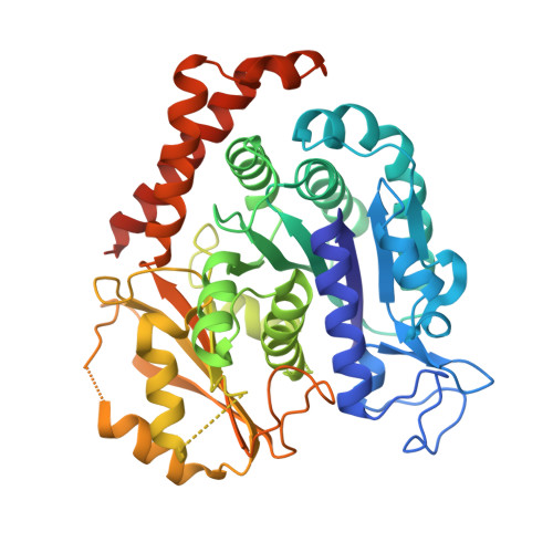
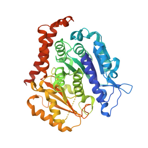
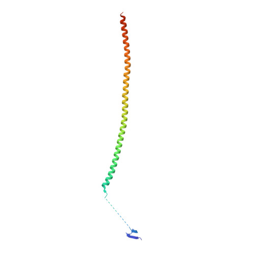
    Targeting the colchicine binding site on tubulin is a promising strategy to develop cancer therapeutics. Herein, we describe our systematic structure-activity relationship studies of benzamide derivatives that lead to an identification of a potent and orally active tubulin inhibitor 48 , which occupied all three zones of the colchicine binding site in the X-ray co-crystal structure, inhibited tubulin polymerization, promoted mitotic blockade and apoptosis, and exhibited significant antiproliferative activities against various cancer cell lines. Compound 48 demonstrated favorable pharmacokinetic profiles, robust in vivo antitumor efficacies, and acceptable safety profiles. Furthermore, 48 overcame drug resistance in the paclitaxel-resistant A549 xenograft model. Collectively, 48 has been advanced into further preclinical evaluation for the development of next-generation microtubule-targeting drugs.


  • Organizational Affiliation

    State Key Laboratory of Bioactive Substance and Function of Natural Medicines, Institute of Materia Medica, Chinese Academy of Medical Sciences and Peking Union Medical College, Beijing 100050, China.


Macromolecules
Find similar proteins by:  (by identity cutoff)  |  3D Structure
Entity ID: 1
MoleculeChains Sequence LengthOrganismDetailsImage
Tubulin alpha-1B chain
A, C
451Sus scrofaMutation(s): 0 
UniProt
Find proteins for Q2XVP4 (Sus scrofa)
Explore Q2XVP4 
Go to UniProtKB:  Q2XVP4
Entity Groups  
Sequence Clusters30% Identity50% Identity70% Identity90% Identity95% Identity100% Identity
UniProt GroupQ2XVP4
Sequence Annotations
Expand
  • Reference Sequence
Find similar proteins by:  (by identity cutoff)  |  3D Structure
Entity ID: 2
MoleculeChains Sequence LengthOrganismDetailsImage
Tubulin beta chain
B, D
445Sus scrofaMutation(s): 0 
UniProt
Find proteins for P02554 (Sus scrofa)
Explore P02554 
Go to UniProtKB:  P02554
Entity Groups  
Sequence Clusters30% Identity50% Identity70% Identity90% Identity95% Identity100% Identity
UniProt GroupP02554
Sequence Annotations
Expand
  • Reference Sequence
Find similar proteins by:  (by identity cutoff)  |  3D Structure
Entity ID: 3
MoleculeChains Sequence LengthOrganismDetailsImage
Stathmin152Sus scrofaMutation(s): 0 
Gene Names: LOC100739720LOC100155126
UniProt
Find proteins for F2Z508 (Sus scrofa)
Explore F2Z508 
Go to UniProtKB:  F2Z508
Entity Groups  
Sequence Clusters30% Identity50% Identity70% Identity90% Identity95% Identity100% Identity
UniProt GroupF2Z508
Sequence Annotations
Expand
  • Reference Sequence
Small Molecules
Ligands 5 Unique
IDChains Name / Formula / InChI Key2D Diagram3D Interactions
GTP
Query on GTP

Download Ideal Coordinates CCD File 
F [auth A],
M [auth C],
P [auth D]
GUANOSINE-5'-TRIPHOSPHATE
C10 H16 N5 O14 P3
XKMLYUALXHKNFT-UUOKFMHZSA-N
GDP
Query on GDP

Download Ideal Coordinates CCD File 
I [auth B]GUANOSINE-5'-DIPHOSPHATE
C10 H15 N5 O11 P2
QGWNDRXFNXRZMB-UUOKFMHZSA-N
IUK (Subject of Investigation/LOI)
Query on IUK

Download Ideal Coordinates CCD File 
K [auth B],
R [auth D]
4-methyl-3-[(4-methylphenyl)sulfonylamino]-~{N}-[(6-methylpyridin-3-yl)methyl]benzamide
C22 H23 N3 O3 S
ZKRBQBKMMRYJKN-UHFFFAOYSA-N
MES
Query on MES

Download Ideal Coordinates CCD File 
J [auth B]2-(N-MORPHOLINO)-ETHANESULFONIC ACID
C6 H13 N O4 S
SXGZJKUKBWWHRA-UHFFFAOYSA-N
MG
Query on MG

Download Ideal Coordinates CCD File 
G [auth A]
H [auth A]
L [auth C]
N [auth C]
O [auth C]
G [auth A],
H [auth A],
L [auth C],
N [auth C],
O [auth C],
Q [auth D]
MAGNESIUM ION
Mg
JLVVSXFLKOJNIY-UHFFFAOYSA-N
Experimental Data & Validation

Experimental Data

  • Method: X-RAY DIFFRACTION
  • Resolution: 2.60 Å
  • R-Value Free: 0.272 
  • R-Value Work: 0.230 
  • R-Value Observed: 0.232 
  • Space Group: P 1 21 1
Unit Cell:
Length ( Å )Angle ( ˚ )
a = 81.87α = 90
b = 102.03β = 106.91
c = 131.13γ = 90
Software Package:
Software NamePurpose
PHENIXrefinement
PHENIXrefinement
xia2data reduction
xia2data scaling
PHENIXphasing

Structure Validation

View Full Validation Report


Ligand Structure Quality Assessment 


Entry History & Funding Information

Deposition Data


Funding OrganizationLocationGrant Number
CAMS Innovation Fund for Medical Sciences (CIFMS)United Kingdom2022-I2M-JB-010

Revision History  (Full details and data files)

  • Version 1.0: 2023-07-26
    Type: Initial release
  • Version 1.1: 2024-02-07
    Changes: Data collection, Database references