7Y65

Cryo-EM structure of C5a peptide-bound C5aR1 in complex with Gi protein


Experimental Data Snapshot

  • Method: ELECTRON MICROSCOPY
  • Resolution: 3.20 Å
  • Aggregation State: PARTICLE 
  • Reconstruction Method: SINGLE PARTICLE 

wwPDB Validation   3D Report Full Report


This is version 1.2 of the entry. See complete history


Literature

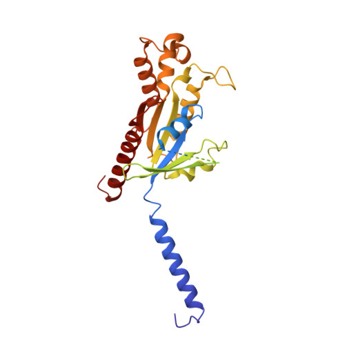
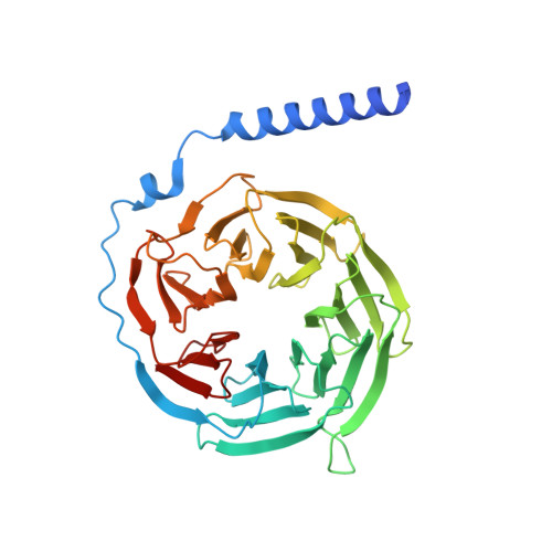
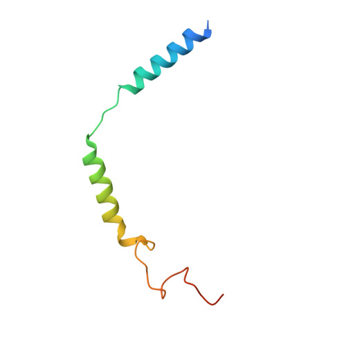
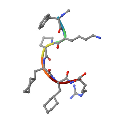
Mechanism of activation and biased signaling in complement receptor C5aR1.

Feng, Y.Zhao, C.Deng, Y.Wang, H.Ma, L.Liu, S.Tian, X.Wang, B.Bin, Y.Chen, P.Yan, W.Fu, P.Shao, Z.

(2023) Cell Res 33: 312-324

  • DOI: https://doi.org/10.1038/s41422-023-00779-2
  • Primary Citation of Related Structures:  
    7Y64, 7Y65, 7Y66, 7Y67

  • PubMed Abstract: 

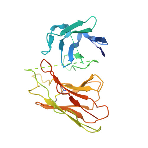
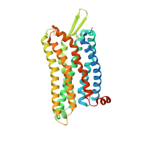
    The complement system plays an important role in the innate immune response to invading pathogens. The complement fragment C5a is one of its important effector components and exerts diverse physiological functions through activation of the C5a receptor 1 (C5aR1) and associated downstream G protein and β-arrestin signaling pathways. Dysfunction of the C5a-C5aR1 axis is linked to numerous inflammatory and immune-mediated diseases, but the structural basis for activation and biased signaling of C5aR1 remains elusive. Here, we present cryo-electron microscopy structures of the activated wild-type C5aR1-G i protein complex bound to each of the following: C5a, the hexapeptidic agonist C5a pep , and the G protein-biased agonist BM213. The structures reveal the landscape of the C5a-C5aR1 interaction as well as a common motif for the recognition of diverse orthosteric ligands. Moreover, combined with mutagenesis studies and cell-based pharmacological assays, we deciphered a framework for biased signaling using different peptide analogs and provided insight into the activation mechanism of C5aR1 by solving the structure of C5aR1 I116A mutant-G i signaling activation complex induced by C089, which exerts antagonism on wild-type C5aR1. In addition, unusual conformational changes in the intracellular end of transmembrane domain 7 and helix 8 upon agonist binding suggest a differential signal transduction process. Collectively, our study provides mechanistic understanding into the ligand recognition, biased signaling modulation, activation, and G i protein coupling of C5aR1, which may facilitate the future design of therapeutic agents.


  • Organizational Affiliation

    Division of Nephrology and Kidney Research Institute, State Key Laboratory of Biotherapy and Cancer Center, West China Hospital, Sichuan University, Chengdu, Sichuan, China.


Macromolecules
Find similar proteins by:  (by identity cutoff)  |  3D Structure
Entity ID: 1
MoleculeChains Sequence LengthOrganismDetailsImage
Guanine nucleotide-binding protein G(i) subunit alpha-1354Homo sapiensMutation(s): 0 
Gene Names: GNAI1
Membrane Entity: Yes 
UniProt & NIH Common Fund Data Resources
Find proteins for P63096 (Homo sapiens)
Explore P63096 
Go to UniProtKB:  P63096
PHAROS:  P63096
GTEx:  ENSG00000127955 
Entity Groups  
Sequence Clusters30% Identity50% Identity70% Identity90% Identity95% Identity100% Identity
UniProt GroupP63096
Sequence Annotations
Expand
  • Reference Sequence
Find similar proteins by:  (by identity cutoff)  |  3D Structure
Entity ID: 2
MoleculeChains Sequence LengthOrganismDetailsImage
Guanine nucleotide-binding protein G(I)/G(S)/G(T) subunit beta-1356Homo sapiensMutation(s): 0 
Gene Names: GNB1
UniProt & NIH Common Fund Data Resources
Find proteins for P62873 (Homo sapiens)
Explore P62873 
Go to UniProtKB:  P62873
PHAROS:  P62873
GTEx:  ENSG00000078369 
Entity Groups  
Sequence Clusters30% Identity50% Identity70% Identity90% Identity95% Identity100% Identity
UniProt GroupP62873
Sequence Annotations
Expand
  • Reference Sequence
Find similar proteins by:  (by identity cutoff)  |  3D Structure
Entity ID: 3
MoleculeChains Sequence LengthOrganismDetailsImage
Guanine nucleotide-binding protein G(I)/G(S)/G(O) subunit gamma-271Homo sapiensMutation(s): 0 
Gene Names: GNG2
Membrane Entity: Yes 
UniProt & NIH Common Fund Data Resources
Find proteins for P59768 (Homo sapiens)
Explore P59768 
Go to UniProtKB:  P59768
PHAROS:  P59768
GTEx:  ENSG00000186469 
Entity Groups  
Sequence Clusters30% Identity50% Identity70% Identity90% Identity95% Identity100% Identity
UniProt GroupP59768
Sequence Annotations
Expand
  • Reference Sequence

Find similar proteins by:  Sequence   |   3D Structure  

Entity ID: 4
MoleculeChains Sequence LengthOrganismDetailsImage
C5apep peptideD [auth L]6synthetic constructMutation(s): 0 
Sequence Annotations
Expand
  • Reference Sequence
Find similar proteins by:  (by identity cutoff)  |  3D Structure
Entity ID: 5
MoleculeChains Sequence LengthOrganismDetailsImage
scFV16E [auth S]267Mus musculusMutation(s): 0 
Entity Groups  
Sequence Clusters30% Identity50% Identity70% Identity90% Identity95% Identity100% Identity
Sequence Annotations
Expand
  • Reference Sequence
Find similar proteins by:  (by identity cutoff)  |  3D Structure
Entity ID: 6
MoleculeChains Sequence LengthOrganismDetailsImage
C5a anaphylatoxin chemotactic receptor 1F [auth D]339Homo sapiensMutation(s): 0 
Gene Names: C5AR1C5ARC5R1
Membrane Entity: Yes 
UniProt & NIH Common Fund Data Resources
Find proteins for P21730 (Homo sapiens)
Explore P21730 
Go to UniProtKB:  P21730
PHAROS:  P21730
GTEx:  ENSG00000197405 
Entity Groups  
Sequence Clusters30% Identity50% Identity70% Identity90% Identity95% Identity100% Identity
UniProt GroupP21730
Sequence Annotations
Expand
  • Reference Sequence
Small Molecules
Modified Residues  2 Unique
IDChains TypeFormula2D DiagramParent
ALC
Query on ALC
D [auth L]L-PEPTIDE LINKINGC9 H17 N O2ALA
MEA
Query on MEA
D [auth L]L-PEPTIDE LINKINGC10 H13 N O2PHE
Experimental Data & Validation

Experimental Data

  • Method: ELECTRON MICROSCOPY
  • Resolution: 3.20 Å
  • Aggregation State: PARTICLE 
  • Reconstruction Method: SINGLE PARTICLE 

Structure Validation

View Full Validation Report


Entry History & Funding Information

Deposition Data


Funding OrganizationLocationGrant Number
National Natural Science Foundation of China (NSFC)China31972916

Revision History  (Full details and data files)

  • Version 1.0: 2023-03-01
    Type: Initial release
  • Version 1.1: 2023-03-08
    Changes: Database references
  • Version 1.2: 2023-04-19
    Changes: Database references