Download Mendeley
Insight into the C-terminal SH3 domain mediated binding of Drosophila Drk to Sos and Dos.
Sayeesh, P.M., Ikeya, T., Sugasawa, H., Watanabe, R., Mishima, M., Inomata, K., Ito, Y.(2022) Biochem Biophys Res Commun 625: 87-93
- PubMed: 35952612 Search on PubMed
- DOI: https://doi.org/10.1016/j.bbrc.2022.08.007
- Primary Citation Related Structures:
7Y4N - PubMed Abstract:
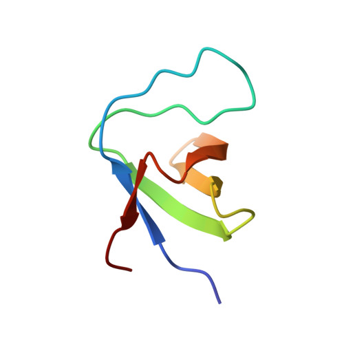
Drk, a Drosophila homologue of human GRB2, interacts with Sevenless (Sev) receptor via its SH2 domain, while the N- and C-terminal SH3 domains (Drk-NSH3 and Drk-CSH3, respectively) are responsible for the interaction with proline-rich motifs (PRMs) of Son of sevenless (Sos) or Daughter of Sevenless (Dos). Drk-NSH3 on its own has a conformational equilibrium between folded and unfolded states, and the folded state is stabilised by the association with a Sos-derived proline-rich peptide with PxxPxR motif. In contrast, Drk-CSH3 is supposed to bind PxxxRxxKP motifs in Dos. Aiming at clarifying the structural and functional differences between the two SH3 domains, we performed NMR studies of Drk-CSH3. The resulting solution structure and the 15 N-relaxation data showed that Drk-CSH3 consists of a stable domain. Large chemical shift perturbation was commonly found around the RT loop and the hydrophobic patch, while there were also changes that occur characteristically for Sos- or Dos-derived peptides. Sos-derived two peptides with PxxPxR motif showed stronger affinity to Drk-CSH3, indicating that the Sos PRMs can bind both N- and C-SH3 domains. Dos-derived two peptides could also bind Drk-CSH3, but with much weaker affinity, suggesting a possibility that any cooperative binding of Dos-PRMs may strengthen the Drk-Dos interaction. The NMR studies as well as the docking simulations provide valuable insights into the biological and biophysical functions of two SH3 domains in Drk.
- Department of Chemistry, Tokyo Metropolitan University, 1-1 Minami-Osawa, Hachioji, Tokyo, 192-0373, Japan.
Organizational Affiliation: