7XU8

Structure of the complex of camel peptidoglycan recognition protein-short (PGRP-S) with heptanoic acid at 2.15 A resolution.


Experimental Data Snapshot

  • Method: X-RAY DIFFRACTION
  • Resolution: 2.15 Å
  • R-Value Free: 0.282 
  • R-Value Work: 0.220 

wwPDB Validation   3D Report Full Report


Ligand Structure Quality Assessment 


This is version 1.2 of the entry. See complete history


Literature

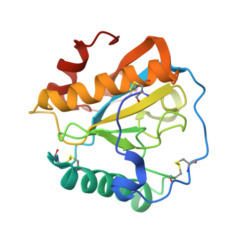
Ligand recognition by peptidoglycan recognition protein-S (PGRP-S): structure of the complex of camel PGRP-S with heptanoic acid at 2.15 angstrom resolution.

Maurya, A.Ahmad, N.Singh, P.K.Viswanathan, V.Kaur, P.Sharma, P.Sharma, S.Singh, T.P.

(2022) Int J Biochem Mol Biol 13: 28-39

  • Primary Citation of Related Structures:  
    7XU8

  • PubMed Abstract: 

    Peptidoglycan recognition proteins (PGRPs) are important components of the innate immune system which provide the first line of defense against invading microbes. There are four members in the family of PGRPs in animals of which PGRP-S is a common domain. It is responsible for the binding to microbial cell wall molecules. In order to understand the mode of binding of PGRP-S to the components of the bacterial cell wall, the structure of the complex of camel PGRP-S (CPGRP-S) with heptanoic acid has been determined at 2.15 Å resolution. The structure determination showed the presence of four crystallographically independent protein molecules which are designated as A, B, C, and D. These four protein molecules associate in the form of two homodimers which are represented as A-B and C-D dimers. The association between molecules A and B gives rise to a shallow cleft on the surface at one end of the dimeric interface. One molecule of heptanoic acid is observed at this binding site in the A-B dimer. The association of C and D molecules results in the formation of a long zig-zag tunnel along with the C-D interface. In the cleft at the C-D interface, three molecules of hydrogen peroxide along with other non-water solvent molecules have been observed. The analysis of the several complexes of CPGRP-S with fatty acids and non-fatty acids such as peptidoglycan, lipopolysaccharide, and lipoteichoic acid shows that the fatty acids bind at the A-B site while non-fatty acids interact through C-D interface.


  • Organizational Affiliation

    Department of Biophysics, All India Institute of Medical Sciences New Delhi, India.


Macromolecules
Find similar proteins by:  (by identity cutoff)  |  3D Structure
Entity ID: 1
MoleculeChains Sequence LengthOrganismDetailsImage
Peptidoglycan recognition protein 1
A, B, C, D
171Camelus dromedariusMutation(s): 0 
UniProt
Find proteins for Q9GK12 (Camelus dromedarius)
Explore Q9GK12 
Go to UniProtKB:  Q9GK12
Entity Groups  
Sequence Clusters30% Identity50% Identity70% Identity90% Identity95% Identity100% Identity
UniProt GroupQ9GK12
Sequence Annotations
Expand
  • Reference Sequence
Small Molecules
Ligands 6 Unique
IDChains Name / Formula / InChI Key2D Diagram3D Interactions
SHV (Subject of Investigation/LOI)
Query on SHV

Download Ideal Coordinates CCD File 
K [auth B]HEPTANOIC ACID
C7 H14 O2
MNWFXJYAOYHMED-UHFFFAOYSA-N
MPD (Subject of Investigation/LOI)
Query on MPD

Download Ideal Coordinates CCD File 
U [auth D](4S)-2-METHYL-2,4-PENTANEDIOL
C6 H14 O2
SVTBMSDMJJWYQN-YFKPBYRVSA-N
EDO (Subject of Investigation/LOI)
Query on EDO

Download Ideal Coordinates CCD File 
E [auth A],
M [auth C],
N [auth C],
R [auth C]
1,2-ETHANEDIOL
C2 H6 O2
LYCAIKOWRPUZTN-UHFFFAOYSA-N
CO3 (Subject of Investigation/LOI)
Query on CO3

Download Ideal Coordinates CCD File 
G [auth A]CARBONATE ION
C O3
BVKZGUZCCUSVTD-UHFFFAOYSA-L
PEO (Subject of Investigation/LOI)
Query on PEO

Download Ideal Coordinates CCD File 
F [auth A]
I [auth B]
J [auth B]
L [auth B]
O [auth C]
F [auth A],
I [auth B],
J [auth B],
L [auth B],
O [auth C],
P [auth C],
Q [auth C],
S [auth D],
T [auth D]
HYDROGEN PEROXIDE
H2 O2
MHAJPDPJQMAIIY-UHFFFAOYSA-N
NA (Subject of Investigation/LOI)
Query on NA

Download Ideal Coordinates CCD File 
H [auth A]SODIUM ION
Na
FKNQFGJONOIPTF-UHFFFAOYSA-N
Experimental Data & Validation

Experimental Data

  • Method: X-RAY DIFFRACTION
  • Resolution: 2.15 Å
  • R-Value Free: 0.282 
  • R-Value Work: 0.220 
  • Space Group: I 2 2 2
Unit Cell:
Length ( Å )Angle ( ˚ )
a = 89.896α = 90
b = 101.637β = 90
c = 163.394γ = 90
Software Package:
Software NamePurpose
REFMACrefinement
DENZOdata reduction
SCALEPACKdata scaling
AMoREphasing

Structure Validation

View Full Validation Report


Ligand Structure Quality Assessment 


Entry History & Funding Information

Deposition Data


Funding OrganizationLocationGrant Number
Not fundedIndia--

Revision History  (Full details and data files)

  • Version 1.0: 2022-06-15
    Type: Initial release
  • Version 1.1: 2022-10-12
    Changes: Database references, Derived calculations
  • Version 1.2: 2023-11-29
    Changes: Data collection, Refinement description