7XRZ

Crystal structure of BRIL and SRP2070_Fab complex


Experimental Data Snapshot

  • Method: X-RAY DIFFRACTION
  • Resolution: 2.10 Å
  • R-Value Free: 0.268 
  • R-Value Work: 0.214 
  • R-Value Observed: 0.216 

wwPDB Validation   3D Report Full Report


This is version 1.2 of the entry. See complete history


Literature

Structural insight into an anti-BRIL Fab as a G-protein-coupled receptor crystallization chaperone.

Miyagi, H.Suzuki, M.Yasunaga, M.Asada, H.Iwata, S.Saito, J.I.

(2023) Acta Crystallogr D Struct Biol 79: 435-441

  • DOI: https://doi.org/10.1107/S205979832300311X
  • Primary Citation of Related Structures:  
    7XRZ

  • PubMed Abstract: 

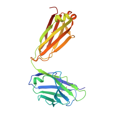
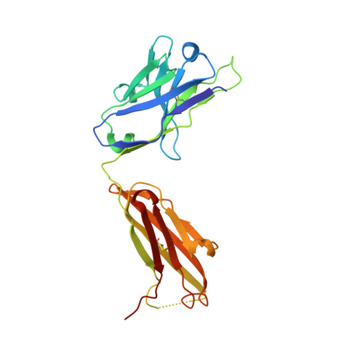
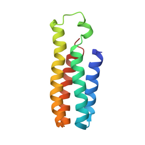
    Structure determination of G-protein-coupled receptors (GPCRs) is key for the successful development of efficient drugs targeting GPCRs. BRIL is a thermostabilized apocytochrome b 562 (with M7W/H102I/R106L mutations) from Escherichia coli and is often used as a GPCR fusion protein for expression and crystallization. SRP2070Fab, an anti-BRIL antibody Fab fragment, has been reported to facilitate and enhance the crystallization of BRIL-fused GPCRs as a crystallization chaperone. This study was conducted to characterize the high-resolution crystal structure of the BRIL-SRP2070Fab complex. The structure of the BRIL-SRP2070Fab complex was determined at 2.1 Å resolution. This high-resolution structure elucidates the binding interaction between BRIL and SRP2070Fab. When binding to BRIL, SRP2070Fab recognizes conformational epitopes, not linear epitopes, on the surface of BRIL helices III and IV, thereby binding perpendicularly to the helices, which indicates stable binding. Additionally, the packing contacts of the BRIL-SRP2070Fab co-crystal are largely due to SRP2070Fab rather than BRIL. The accumulation of SRP2070Fab molecules by stacking is remarkable and is consistent with the finding that stacking of SRP2070Fab is predominant in known crystal structures of BRIL-fused GPCRs complexed with SRP2070Fab. These findings clarified the mechanism of SRP2070Fab as a crystallization chaperone. Moreover, these data will be useful in the structure-based drug design of membrane-protein drug targets.


  • Organizational Affiliation

    R&D Division, Kyowa Kirin Co. Ltd, Tokyo, Japan.


Macromolecules
Find similar proteins by:  (by identity cutoff)  |  3D Structure
Entity ID: 1
MoleculeChains Sequence LengthOrganismDetailsImage
IGG LIGHT CHAINA [auth L],
D [auth A]
214Mus musculusMutation(s): 0 
Entity Groups  
Sequence Clusters30% Identity50% Identity70% Identity90% Identity95% Identity100% Identity
Sequence Annotations
Expand
  • Reference Sequence
Find similar proteins by:  (by identity cutoff)  |  3D Structure
Entity ID: 2
MoleculeChains Sequence LengthOrganismDetailsImage
IGG HEAVY CHAINB [auth H],
E [auth B]
221Mus musculusMutation(s): 0 
Entity Groups  
Sequence Clusters30% Identity50% Identity70% Identity90% Identity95% Identity100% Identity
Sequence Annotations
Expand
  • Reference Sequence
Find similar proteins by:  (by identity cutoff)  |  3D Structure
Entity ID: 3
MoleculeChains Sequence LengthOrganismDetailsImage
Soluble cytochrome b562C [auth X],
F [auth Y]
106Escherichia coliMutation(s): 3 
Gene Names: cybC
UniProt
Find proteins for P0ABE7 (Escherichia coli)
Explore P0ABE7 
Go to UniProtKB:  P0ABE7
Entity Groups  
Sequence Clusters30% Identity50% Identity70% Identity90% Identity95% Identity100% Identity
UniProt GroupP0ABE7
Sequence Annotations
Expand
  • Reference Sequence
Experimental Data & Validation

Experimental Data

  • Method: X-RAY DIFFRACTION
  • Resolution: 2.10 Å
  • R-Value Free: 0.268 
  • R-Value Work: 0.214 
  • R-Value Observed: 0.216 
  • Space Group: C 1 2 1
Unit Cell:
Length ( Å )Angle ( ˚ )
a = 93.828α = 90
b = 75.239β = 99.67
c = 152.231γ = 90
Software Package:
Software NamePurpose
Aimlessdata scaling
PHASERphasing
REFMACrefinement
PDB_EXTRACTdata extraction
XDSdata reduction

Structure Validation

View Full Validation Report


Entry History & Funding Information

Deposition Data


Funding OrganizationLocationGrant Number
Not funded--

Revision History  (Full details and data files)

  • Version 1.0: 2023-05-10
    Type: Initial release
  • Version 1.1: 2023-05-31
    Changes: Database references
  • Version 1.2: 2023-11-29
    Changes: Data collection, Refinement description