Structural mechanism of a dual-functional enzyme DgpA/B/C as both a C -glycoside cleaving enzyme and an O - to C -glycoside isomerase.
He, P., Wang, S., Li, S., Liu, S., Zhou, S., Wang, J., Tao, J., Wang, D., Wang, R., Ma, W.(2023) Acta Pharm Sin B 13: 246-255
- PubMed: 36815035 Search on PubMedSearch on PubMed Central
- DOI: https://doi.org/10.1016/j.apsb.2022.05.022
- Primary Citation Related Structures:
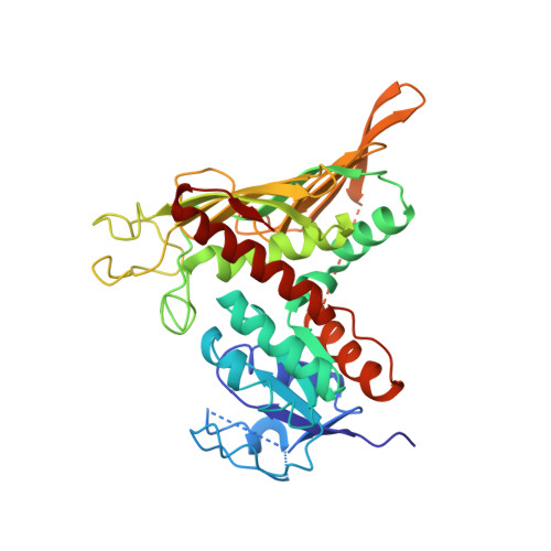
7XR9, 7XRE, 7XRF - PubMed Abstract:
The C -glycosidic bond that connects the sugar moiety with aglycone is difficult to be broken or made due to its inert nature. The knowledge of C -glycoside breakdown and synthesis is very limited. Recently, the enzyme DgpA/B/C cascade from a human intestinal bacterium PUE was identified to specifically cleave the C -glycosidic bond of puerarin (daidzein-8- C -glucoside). Here we investigated how puerarin is recognized and oxidized by DgpA based on crystal structures of DgpA with or without substrate and biochemical characterization. More strikingly, we found that apart from being a C -glycoside cleaving enzyme, DgpA/B/C is capable of efficiently converting O - to C -glycoside showing the activity as a structure isomerase. A possible mechanistic model was proposed dependently of the simulated complex structure of DgpB/C with 3″-oxo-daidzin and structure-based mutagenesis. Our findings not only shed light on understanding the enzyme-mediated C -glycosidic bond breakage and formation, but also may help to facilitate stereospecific C -glycoside synthesis in pharmaceutical industry.
- School of Life Science, Beijing University of Chinese Medicine, Beijing 102488, China.
Organizational Affiliation: