7XML

Cryo-EM structure of PEIP-Bs_enolase complex


Experimental Data Snapshot

  • Method: ELECTRON MICROSCOPY
  • Resolution: 3.20 Å
  • Aggregation State: PARTICLE 
  • Reconstruction Method: SINGLE PARTICLE 

wwPDB Validation   3D Report Full Report


This is version 1.0 of the entry. See complete history


Literature

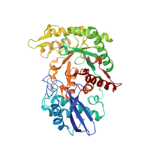
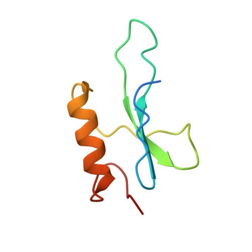
Bacteriophage protein PEIP is a potent Bacillus subtilis enolase inhibitor.

Zhang, K.Li, S.Wang, Y.Wang, Z.Mulvenna, N.Yang, H.Zhang, P.Chen, H.Li, Y.Wang, H.Gao, Y.Wigneshweraraj, S.Matthews, S.Zhang, K.Liu, B.

(2022) Cell Rep 40: 111026-111026

  • DOI: https://doi.org/10.1016/j.celrep.2022.111026
  • Primary Citation of Related Structures:  
    7XML

  • PubMed Abstract: 

    Enolase is a highly conserved enzyme that presents in all organisms capable of glycolysis or fermentation. Its immediate product phosphoenolpyruvate is essential for other important processes like peptidoglycan synthesis and the phosphotransferase system in bacteria. Therefore, enolase inhibitors are of great interest. Here, we report that Gp60, a phage-encoded enolase inhibitor protein (PEIP) of bacteriophage SPO1 for Bacillus subtilis, is an enolase inhibitor. PEIP-expressing bacteria exhibit growth attenuation, thinner cell walls, and safranin color in Gram staining owing to impaired peptidoglycan synthesis. We solve the structure of PEIP-enolase tetramer and show that PEIP disassembles enolase by disrupting the basic dimer unit. The structure reveals that PEIP does not compete for substrate binding but induces a cascade of conformational changes that limit accessibility to the enolase catalytic site. This phage-inspired disassembly of enolase represents an alternative strategy for the development of anti-microbial drugs.


  • Organizational Affiliation

    BioBank, The First Affiliated Hospital of Xi'an Jiaotong University, Shaanxi 710061, China; Institute for Protein Science and Phage Research, The First Affiliated Hospital of Xi'an Jiaotong University, Shaanxi 710061, China.


Macromolecules
Find similar proteins by:  (by identity cutoff)  |  3D Structure
Entity ID: 1
MoleculeChains Sequence LengthOrganismDetailsImage
EnolaseA,
C [auth B]
430Bacillus subtilis subsp. subtilis str. 168Mutation(s): 0 
Gene Names: enoBSU33900
EC: 4.2.1.11
UniProt
Find proteins for P37869 (Bacillus subtilis (strain 168))
Explore P37869 
Go to UniProtKB:  P37869
Entity Groups  
Sequence Clusters30% Identity50% Identity70% Identity90% Identity95% Identity100% Identity
UniProt GroupP37869
Sequence Annotations
Expand
  • Reference Sequence
Find similar proteins by:  (by identity cutoff)  |  3D Structure
Entity ID: 2
MoleculeChains Sequence LengthOrganismDetailsImage
Putative gene 60 proteinB [auth C],
D
73Okubovirus SPO1Mutation(s): 0 
Gene Names: 60
UniProt
Find proteins for O48414 (Bacillus phage SP01)
Explore O48414 
Go to UniProtKB:  O48414
Entity Groups  
Sequence Clusters30% Identity50% Identity70% Identity90% Identity95% Identity100% Identity
UniProt GroupO48414
Sequence Annotations
Expand
  • Reference Sequence
Experimental Data & Validation

Experimental Data

  • Method: ELECTRON MICROSCOPY
  • Resolution: 3.20 Å
  • Aggregation State: PARTICLE 
  • Reconstruction Method: SINGLE PARTICLE 

Structure Validation

View Full Validation Report


Entry History & Funding Information

Deposition Data


Funding OrganizationLocationGrant Number
Not funded--

Revision History  (Full details and data files)

  • Version 1.0: 2022-07-27
    Type: Initial release