7V62

Crystal structure of human OSBP ORD in complex with cholesterol


Experimental Data Snapshot

  • Method: X-RAY DIFFRACTION
  • Resolution: 3.25 Å
  • R-Value Free: 0.252 
  • R-Value Work: 0.213 
  • R-Value Observed: 0.215 

wwPDB Validation   3D Report Full Report


Ligand Structure Quality Assessment 


This is version 1.2 of the entry. See complete history


Literature

Ligand Recognition by the Lipid Transfer Domain of Human OSBP Is Important for Enterovirus Replication.

Kobayashi, J.Arita, M.Sakai, S.Kojima, H.Senda, M.Senda, T.Hanada, K.Kato, R.

(2022) ACS Infect Dis 8: 1161-1170

  • DOI: https://doi.org/10.1021/acsinfecdis.2c00108
  • Primary Citation of Related Structures:  
    7V62

  • PubMed Abstract: 

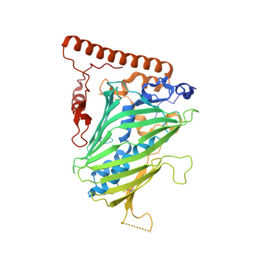
    Oxysterol-binding protein (OSBP), which transports cholesterol and phosphatidylinositol 4-monophosphate (PtdIns[4]P) between different organelles, serves as a conserved host factor for the replication of various viruses, and OSBP inhibitors exhibit antiviral effects. Here, we determined the crystal structure of the lipid transfer domain of human OSBP in complex with endogenous cholesterol. The hydrocarbon tail and tetracyclic ring of cholesterol interact with the hydrophobic tunnel of OSBP, and the hydroxyl group of cholesterol forms a hydrogen bond network at the bottom of the tunnel. Systematic mutagenesis of the ligand-binding region revealed that M446W and L590W substitutions confer functional tolerance to an OSBP inhibitor, T-00127-HEV2. Employing the M446W variant as a functional replacement for the endogenous OSBP in the presence of T-00127-HEV2, we have identified previously unappreciated amino acid residues required for viral replication. The combined use of the inhibitor and the OSBP variant will be useful in elucidating the enigmatic in vivo functions of OSBP.


  • Organizational Affiliation

    Structural Biology Research Center, Institute of Materials Structure Science, High Energy Accelerator Research Organization (KEK), 1-1 Oho, Tsukuba, Ibaraki 305-0801, Japan.


Macromolecules
Find similar proteins by:  (by identity cutoff)  |  3D Structure
Entity ID: 1
MoleculeChains Sequence LengthOrganismDetailsImage
Oxysterol-binding protein 1
A, B, C, D
404Homo sapiensMutation(s): 0 
Gene Names: OSBPOSBP1
UniProt & NIH Common Fund Data Resources
Find proteins for P22059 (Homo sapiens)
Explore P22059 
Go to UniProtKB:  P22059
PHAROS:  P22059
GTEx:  ENSG00000110048 
Entity Groups  
Sequence Clusters30% Identity50% Identity70% Identity90% Identity95% Identity100% Identity
UniProt GroupP22059
Sequence Annotations
Expand
  • Reference Sequence
Small Molecules
Ligands 4 Unique
IDChains Name / Formula / InChI Key2D Diagram3D Interactions
CLR (Subject of Investigation/LOI)
Query on CLR

Download Ideal Coordinates CCD File 
E [auth A],
J [auth B],
M [auth C],
Q [auth D]
CHOLESTEROL
C27 H46 O
HVYWMOMLDIMFJA-DPAQBDIFSA-N
CIT
Query on CIT

Download Ideal Coordinates CCD File 
H [auth A]CITRIC ACID
C6 H8 O7
KRKNYBCHXYNGOX-UHFFFAOYSA-N
DTT
Query on DTT

Download Ideal Coordinates CCD File 
F [auth A]
G [auth A]
K [auth B]
N [auth C]
O [auth C]
F [auth A],
G [auth A],
K [auth B],
N [auth C],
O [auth C],
R [auth D]
2,3-DIHYDROXY-1,4-DITHIOBUTANE
C4 H10 O2 S2
VHJLVAABSRFDPM-IMJSIDKUSA-N
DMS
Query on DMS

Download Ideal Coordinates CCD File 
I [auth A],
L [auth C],
P [auth C],
S [auth D]
DIMETHYL SULFOXIDE
C2 H6 O S
IAZDPXIOMUYVGZ-UHFFFAOYSA-N
Experimental Data & Validation

Experimental Data

  • Method: X-RAY DIFFRACTION
  • Resolution: 3.25 Å
  • R-Value Free: 0.252 
  • R-Value Work: 0.213 
  • R-Value Observed: 0.215 
  • Space Group: P 43 21 2
Unit Cell:
Length ( Å )Angle ( ˚ )
a = 111.88α = 90
b = 111.88β = 90
c = 367.88γ = 90
Software Package:
Software NamePurpose
PHENIXrefinement
XDSdata reduction
XDSdata scaling
CRANK2phasing

Structure Validation

View Full Validation Report


Ligand Structure Quality Assessment 


Entry History & Funding Information

Deposition Data


Funding OrganizationLocationGrant Number
Japan Agency for Medical Research and Development (AMED)Japan21am0101083j0005

Revision History  (Full details and data files)

  • Version 1.0: 2022-06-08
    Type: Initial release
  • Version 1.1: 2022-06-22
    Changes: Database references
  • Version 1.2: 2023-11-29
    Changes: Data collection, Refinement description