7UYI | pdb_00007uyi

Crystal structure of the computationally optimized broadly reactive H1 influenza hemagglutinin P1


Experimental Data Snapshot

  • Method: X-RAY DIFFRACTION
  • Resolution: 3.00 Å
  • R-Value Free: 
    0.270 (Depositor), 0.275 (DCC) 
  • R-Value Work: 
    0.226 (Depositor) 
  • R-Value Observed: 
    0.228 (Depositor) 

wwPDB Validation 3D Report Full Report

Validation slider image for 7UYI

Ligand Structure Quality Assessment 


This is version 1.3 of the entry. See complete history

Literature

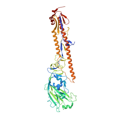
Structural insights into the broad protection against H1 influenza viruses by a computationally optimized hemagglutinin vaccine.

Dzimianski, J.V.Han, J.Sautto, G.A.O'Rourke, S.M.Cruz, J.M.Pierce, S.R.Ecker, J.W.Carlock, M.A.Nagashima, K.A.Mousa, J.J.Ross, T.M.Ward, A.B.DuBois, R.M.

(2023) Commun Biol 6: 454-454

  • DOI: https://doi.org/10.1038/s42003-023-04793-3
  • Primary Citation Related Structures: 
    7UYI, 8CT6

  • PubMed Abstract: 

    Influenza virus poses an ongoing human health threat with pandemic potential. Due to mutations in circulating strains, formulating effective vaccines remains a challenge. The use of computationally optimized broadly reactive antigen (COBRA) hemagglutinin (HA) proteins is a promising vaccine strategy to protect against a wide range of current and future influenza viruses. Though effective in preclinical studies, the mechanistic basis driving the broad reactivity of COBRA proteins remains to be elucidated. Here, we report the crystal structure of the COBRA HA termed P1 and identify antigenic and glycosylation properties that contribute to its immunogenicity. We further report the cryo-EM structure of the P1-elicited broadly neutralizing antibody 1F8 bound to COBRA P1, revealing 1F8 to recognize an atypical receptor binding site epitope via an unexpected mode of binding.


  • Organizational Affiliation
    • Department of Biomolecular Engineering, University of California Santa Cruz, Santa Cruz, CA, USA.

Macromolecule Content 

  • Total Structure Weight: 356.48 kDa 
  • Atom Count: 23,338 
  • Modeled Residue Count: 2,852 
  • Deposited Residue Count: 3,078 
  • Unique protein chains: 1

Macromolecules

Find similar proteins by:|  3D Structure
Entity ID: 1
MoleculeChains  Sequence LengthOrganismDetailsImage
COBRA P1 HA513Influenza A virusMutation(s): 0 

Oligosaccharides

Help  
Entity ID: 2
MoleculeChains Length2D Diagram GlycosylationD Interactions
alpha-D-mannopyranose-(1-3)-beta-D-mannopyranose-(1-4)-2-acetamido-2-deoxy-beta-D-glucopyranose-(1-4)-2-acetamido-2-deoxy-beta-D-glucopyranose
G, P
4N/AN-Glycosylation
Glycosylation Resources
GlyTouCan: G81315DD
GlyCosmos: G81315DD
GlyGen: G81315DD
Entity ID: 3
MoleculeChains Length2D Diagram GlycosylationD Interactions
2-acetamido-2-deoxy-beta-D-glucopyranose-(1-4)-2-acetamido-2-deoxy-beta-D-glucopyranose
H, I, K, O, Q
2N/AN-Glycosylation
Glycosylation Resources
GlyTouCan: G42666HT
GlyCosmos: G42666HT
GlyGen: G42666HT
Entity ID: 4
MoleculeChains Length2D Diagram GlycosylationD Interactions
beta-D-mannopyranose-(1-4)-2-acetamido-2-deoxy-beta-D-glucopyranose-(1-4)-2-acetamido-2-deoxy-beta-D-glucopyranose
J, L, M
3N/AN-Glycosylation
Glycosylation Resources
GlyTouCan: G15407YE
GlyCosmos: G15407YE
GlyGen: G15407YE
Entity ID: 5
MoleculeChains Length2D Diagram GlycosylationD Interactions
alpha-D-mannopyranose-(1-3)-[alpha-D-mannopyranose-(1-6)]beta-D-mannopyranose-(1-4)-2-acetamido-2-deoxy-beta-D-glucopyranose-(1-4)-2-acetamido-2-deoxy-beta-D-glucopyranose
N, R
5N/AN-Glycosylation
Glycosylation Resources
GlyTouCan: G22768VO
GlyCosmos: G22768VO
GlyGen: G22768VO

Small Molecules

Ligands 1 Unique
IDChains Name / Formula / InChI Key2D Diagram3D Interactions
NAG

Query on NAG


Download:Ideal Coordinates CCD File
AA [auth C]
BA [auth C]
CA [auth C]
DA [auth C]
EA [auth E]
AA [auth C],
BA [auth C],
CA [auth C],
DA [auth C],
EA [auth E],
FA [auth F],
GA [auth F],
HA [auth D],
IA [auth D],
JA [auth D],
KA [auth D],
S [auth A],
T [auth A],
U [auth A],
V [auth A],
W [auth B],
X [auth B],
Y [auth B],
Z [auth B]
2-acetamido-2-deoxy-beta-D-glucopyranose
C8 H15 N O6
OVRNDRQMDRJTHS-FMDGEEDCSA-N

Experimental Data & Validation

Experimental Data

  • Method: X-RAY DIFFRACTION
  • Resolution: 3.00 Å
  • R-Value Free:  0.270 (Depositor), 0.275 (DCC) 
  • R-Value Work:  0.226 (Depositor) 
  • R-Value Observed: 0.228 (Depositor) 
Space Group: C 1 2 1
Unit Cell:
Length ( Å )Angle ( ˚ )
a = 264.849α = 90
b = 77.56β = 93.773
c = 222.523γ = 90
Software Package:
Software NamePurpose
Blu-Icedata collection
DIALSdata reduction
Aimlessdata scaling
PHASERphasing
PHENIXrefinement
Cootmodel building

Structure Validation

View Full Validation Report


Ligand Structure Quality Assessment 


Entry History 

& Funding Information

Deposition Data


Funding OrganizationLocationGrant Number
National Institutes of Health/National Institute Of Allergy and Infectious Diseases (NIH/NIAID)United States75N93019C00052

Revision History  (Full details and data files)

  • Version 1.0: 2023-04-19
    Type: Initial release
  • Version 1.1: 2023-05-03
    Changes: Database references
  • Version 1.2: 2024-05-01
    Changes: Data collection, Database references
  • Version 1.3: 2024-10-09
    Changes: Structure summary