7UQJ

Cryo-EM structure of the S. cerevisiae chromatin remodeler Yta7 hexamer bound to ATPgS and histone H3 tail in state II


Experimental Data Snapshot

  • Method: ELECTRON MICROSCOPY
  • Resolution: 3.00 Å
  • Aggregation State: PARTICLE 
  • Reconstruction Method: SINGLE PARTICLE 

wwPDB Validation   3D Report Full Report


This is version 1.1 of the entry. See complete history


Literature

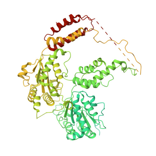
The Saccharomyces cerevisiae Yta7 ATPase hexamer contains a unique bromodomain tier that functions in nucleosome disassembly.

Wang, F.Feng, X.He, Q.Li, H.Li, H.

(2022) J Biol Chem 299: 102852-102852

  • DOI: https://doi.org/10.1016/j.jbc.2022.102852
  • Primary Citation of Related Structures:  
    7UQI, 7UQJ, 7UQK

  • PubMed Abstract: 

    The Saccharomyces cerevisiae Yta7 is a chromatin remodeler harboring a histone-interacting bromodomain (BRD) and two AAA+ modules. It is not well understood how Yta7 recognizes the histone H3 tail to promote nucleosome disassembly for DNA replication or RNA transcription. By cryo-EM analysis, here we show that Yta7 assembles a three-tiered hexamer with a top BRD tier, a middle AAA1 tier, and a bottom AAA2 tier. Unexpectedly, the Yta7 BRD stabilizes a four-stranded β-helix, termed BRD-interacting motif (BIM), of the largely disordered N-terminal region. The BIM motif is unique to the baker's yeast, and we show both BRD and BIM contribute to nucleosome recognition. We found that Yta7 binds both acetylated and nonacetylated H3 peptides but with a higher affinity for the unmodified peptide. This property is consistent with the absence of key residues of canonical BRDs involved in acetylated peptide recognition and the role of Yta7 in general nucleosome remodeling. Interestingly, the BRD tier exists in a spiral and a flat-ring form on top of the Yta7 AAA+ hexamer. The spiral is likely in a nucleosome-searching mode because the bottom BRD blocks the entry to the AAA+ chamber. The flat ring may be in a nucleosome disassembly state because the entry is unblocked and the H3 peptide has entered the AAA+ chamber and is stabilized by the AAA1 pore loops 1 and 2. Indeed, we show that the BRD tier is a flat ring when bound to the nucleosome. Overall, our study sheds light on the nucleosome disassembly by Yta7.


  • Organizational Affiliation

    Department of Structural Biology, Van Andel Institute, Grand Rapids, Michigan, USA.


Macromolecules
Find similar proteins by:  (by identity cutoff)  |  3D Structure
Entity ID: 1
MoleculeChains Sequence LengthOrganismDetailsImage
ATPase histone chaperone YTA7
A, B, C, D, E
A, B, C, D, E, F
1,416Saccharomyces cerevisiaeMutation(s): 0 
Gene Names: YTA7YGR270W
EC: 3.6.1
UniProt
Find proteins for P40340 (Saccharomyces cerevisiae (strain ATCC 204508 / S288c))
Explore P40340 
Go to UniProtKB:  P40340
Entity Groups  
Sequence Clusters30% Identity50% Identity70% Identity90% Identity95% Identity100% Identity
UniProt GroupP40340
Sequence Annotations
Expand
  • Reference Sequence

Find similar proteins by:  Sequence   |   3D Structure  

Entity ID: 2
MoleculeChains Sequence LengthOrganismDetailsImage
Histone H325Saccharomyces cerevisiaeMutation(s): 0 
UniProt
Find proteins for P61830 (Saccharomyces cerevisiae (strain ATCC 204508 / S288c))
Explore P61830 
Go to UniProtKB:  P61830
Entity Groups  
Sequence Clusters30% Identity50% Identity70% Identity90% Identity95% Identity100% Identity
UniProt GroupP61830
Sequence Annotations
Expand
  • Reference Sequence
Small Molecules
Ligands 3 Unique
IDChains Name / Formula / InChI Key2D Diagram3D Interactions
AGS (Subject of Investigation/LOI)
Query on AGS

Download Ideal Coordinates CCD File 
I [auth B],
J [auth C],
L [auth D],
N [auth E],
P [auth F]
PHOSPHOTHIOPHOSPHORIC ACID-ADENYLATE ESTER
C10 H16 N5 O12 P3 S
NLTUCYMLOPLUHL-KQYNXXCUSA-N
ADP (Subject of Investigation/LOI)
Query on ADP

Download Ideal Coordinates CCD File 
H [auth A]ADENOSINE-5'-DIPHOSPHATE
C10 H15 N5 O10 P2
XTWYTFMLZFPYCI-KQYNXXCUSA-N
MG (Subject of Investigation/LOI)
Query on MG

Download Ideal Coordinates CCD File 
K [auth C],
M [auth D],
O [auth E],
Q [auth F]
MAGNESIUM ION
Mg
JLVVSXFLKOJNIY-UHFFFAOYSA-N
Experimental Data & Validation

Experimental Data

  • Method: ELECTRON MICROSCOPY
  • Resolution: 3.00 Å
  • Aggregation State: PARTICLE 
  • Reconstruction Method: SINGLE PARTICLE 

Structure Validation

View Full Validation Report


Entry History & Funding Information

Deposition Data


Funding OrganizationLocationGrant Number
National Institutes of Health/National Institute of General Medical Sciences (NIH/NIGMS)United StatesGM131754

Revision History  (Full details and data files)

  • Version 1.0: 2023-02-01
    Type: Initial release
  • Version 1.1: 2023-02-08
    Changes: Database references