Differentiating the roles of Mycobacterium tuberculosis substrate binding proteins, FecB and FecB2, in iron uptake.
de Miranda, R., Cuthbert, B.J., Klevorn, T., Chao, A., Mendoza, J., Arbing, M., Sieminski, P.J., Papavinasasundaram, K., Abdul-Hafiz, S., Chan, S., Sassetti, C.M., Ehrt, S., Goulding, C.W.(2023) PLoS Pathog 19: e1011650-e1011650
- PubMed: 37747938 Search on PubMedSearch on PubMed Central
- DOI: https://doi.org/10.1371/journal.ppat.1011650
- Primary Citation Related Structures:
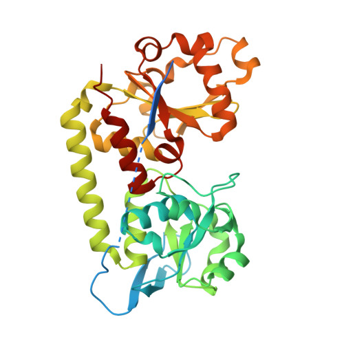
7UQ0 - PubMed Abstract:
Mycobacterium tuberculosis (Mtb), the causative agent of tuberculosis, poses a great threat to human health. With the emergence of drug resistant Mtb strains, new therapeutics are desperately needed. As iron is critical to the growth and survival of Mtb, mechanisms through which Mtb acquires host iron represent attractive therapeutic targets. Mtb scavenges host iron via Mtb siderophore-dependent and heme iron uptake pathways. While multiple studies describe the import of heme and ferric-siderophores and the export of apo-siderophores across the inner membrane, little is known about their transport across the periplasm and cell-wall environments. Mtb FecB and FecB2 are predicted periplasmic binding proteins implicated in host iron acquisition; however, their precise roles are not well understood. This study sought to differentiate the roles FecB and FecB2 play in Mtb iron acquisition. The crystallographic structures of Mtb FecB and FecB2 were determined to 2.0 Å and 2.2 Å resolution, respectively, and show distinct ligand binding pockets. In vitro ligand binding experiments for FecB and FecB2 were performed with heme and bacterial siderophores from Mtb and other species, revealing that both FecB and FecB2 bind heme, while only FecB binds the Mtb sideophore ferric-carboxymycobactin (Fe-cMB). Subsequent structure-guided mutagenesis of FecB identified a single glutamate residue-Glu339-that significantly contributes to Fe-cMB binding. A role for FecB in the Mtb siderophore-mediated iron acquisition pathway was corroborated by Mycobacterium smegmatis and Mtb pull-down assays, which revealed interactions between FecB and members of the mycobacterial siderophore export and import machinery. Similarly, pull-down assays with FecB2 confirms its role in heme uptake revealing interactions with a potential inner membrane heme importer. Due to ligand preference and protein partners, our data suggest that Mtb FecB plays a role in siderophore-dependent iron and heme acquisition pathways; in addition, we confirm that Mtb FecB2 is involved in heme uptake.
- Department of Molecular Biology & Biochemistry, University of California Irvine, Irvine, California, United States of America.
Organizational Affiliation: