7UKC | pdb_00007ukc

Human Kv4.2-KChIP2 channel complex in an inactivated state, class 1, transmembrane region


Experimental Data Snapshot

  • Method: ELECTRON MICROSCOPY
  • Resolution: 3.00 Å
  • Aggregation State: PARTICLE 
  • Reconstruction Method: SINGLE PARTICLE 

wwPDB Validation 3D Report Full Report

Validation slider image for 7UKC

This is version 1.2 of the entry. See complete history

Literature

Activation and closed-state inactivation mechanisms of the human voltage-gated K V 4 channel complexes.

Ye, W.Zhao, H.Dai, Y.Wang, Y.Lo, Y.H.Jan, L.Y.Lee, C.H.

(2022) Mol Cell 82: 2427

  • DOI: https://doi.org/10.1016/j.molcel.2022.04.032
  • Primary Citation Related Structures: 
    7UK5, 7UKC, 7UKD, 7UKE, 7UKF, 7UKG, 7UKH

  • PubMed Abstract: 

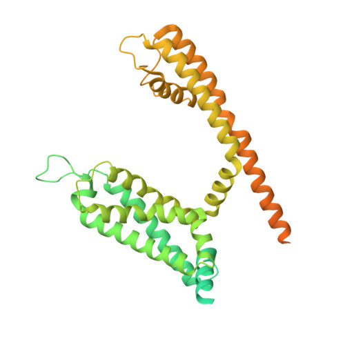
    The voltage-gated ion channel activity depends on both activation (transition from the resting state to the open state) and inactivation. Inactivation is a self-restraint mechanism to limit ion conduction and is as crucial to membrane excitability as activation. Inactivation can occur when the channel is open or closed. Although open-state inactivation is well understood, the molecular basis of closed-state inactivation has remained elusive. We report cryo-EM structures of human K V 4.2 channel complexes in inactivated, open, and closed states. Closed-state inactivation of K V 4 involves an unprecedented symmetry breakdown for pore closure by only two of the four S4-S5 linkers, distinct from known mechanisms of open-state inactivation. We further capture K V 4 in a putative resting state, revealing how voltage sensor movements control the pore. Moreover, our structures provide insights regarding channel modulation by KChIP2 and DPP6 auxiliary subunits. Our findings elucidate mechanisms of closed-state inactivation and voltage-dependent activation of the K V 4 channel.


  • Organizational Affiliation
    • Department of Physiology, University of California, San Francisco, CA 94158, USA.

Macromolecule Content 

  • Total Structure Weight: 236.35 kDa 
  • Atom Count: 8,000 
  • Modeled Residue Count: 1,036 
  • Deposited Residue Count: 2,096 
  • Unique protein chains: 1

Macromolecules

Find similar proteins by:|  3D Structure
Entity ID: 1
MoleculeChains  Sequence LengthOrganismDetailsImage
Potassium voltage-gated channel subfamily D member 2A [auth B],
B [auth C],
C [auth D],
D [auth A]
524Homo sapiensMutation(s): 0 
Gene Names: KCND2KIAA1044
Membrane Entity: Yes 
UniProt & NIH Common Fund Data Resources
Find proteins for Q9NZV8 (Homo sapiens)
Explore Q9NZV8 
Go to UniProtKB:  Q9NZV8
PHAROS:  Q9NZV8
GTEx:  ENSG00000184408 
Entity Groups
Sequence Clusters30% Identity50% Identity70% Identity90% Identity95% Identity100% Identity
UniProt GroupQ9NZV8
Sequence Annotations
Expand
Reference Sequence

Experimental Data & Validation

Experimental Data

  • Method: ELECTRON MICROSCOPY
  • Resolution: 3.00 Å
  • Aggregation State: PARTICLE 
  • Reconstruction Method: SINGLE PARTICLE 

Structure Validation

View Full Validation Report


Entry History 

& Funding Information

Deposition Data


Funding OrganizationLocationGrant Number
National Institutes of Health/National Institute of General Medical Sciences (NIH/NIGMS)United StatesGM143282

Revision History  (Full details and data files)

  • Version 1.0: 2022-06-29
    Type: Initial release
  • Version 1.1: 2022-07-20
    Changes: Database references
  • Version 1.2: 2024-02-14
    Changes: Data collection