7SEC

Crystal structure of human Fibrillarin in complex with compound 1a


Experimental Data Snapshot

  • Method: X-RAY DIFFRACTION
  • Resolution: 1.90 Å
  • R-Value Free: 0.266 
  • R-Value Work: 0.223 
  • R-Value Observed: 0.225 

wwPDB Validation   3D Report Full Report


Ligand Structure Quality Assessment 


This is version 1.1 of the entry. See complete history


Literature

Discovery of Cofactor Competitive Inhibitors against the Human Methyltransferase Fibrillarin.

Shi, Y.El-Deeb, I.M.Masic, V.Hartley-Tassell, L.Maggioni, A.Itzstein, M.V.Ve, T.

(2021) Pharmaceuticals (Basel) 15

  • DOI: https://doi.org/10.3390/ph15010026
  • Primary Citation of Related Structures:  
    7SE6, 7SE7, 7SE8, 7SE9, 7SEA, 7SEB, 7SEC, 7SED

  • PubMed Abstract: 

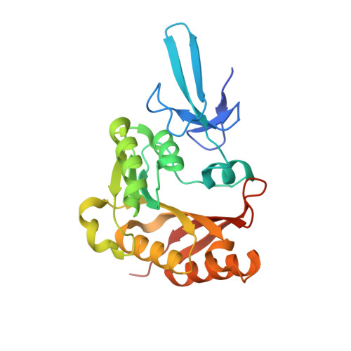
    Fibrillarin (FBL) is an essential and evolutionarily highly conserved S-adenosyl methionine (SAM) dependent methyltransferase. It is the catalytic component of a multiprotein complex that facilitates 2'- O -methylation of ribosomal RNAs (rRNAs), a modification essential for accurate and efficient protein synthesis in eukaryotic cells. It was recently established that human FBL (hFBL) is critical for Nipah, Hendra, and respiratory syncytial virus infections. In addition, overexpression of hFBL contributes towards tumorgenesis and is associated with poor survival in patients with breast cancer, suggesting that hFBL is a potential target for the development of both antiviral and anticancer drugs. An attractive strategy to target cofactor-dependent enzymes is the selective inhibition of cofactor binding, which has been successful for the development of inhibitors against several protein methyltransferases including PRMT5, DOT1L, and EZH2. In this work, we solved crystal structures of the methyltransferase domain of hFBL in apo form and in complex with the cofactor SAM. Screening of a fluorinated fragment library, via X-ray crystallography and 19F NMR spectroscopy, yielded seven hit compounds that competed with cofactor binding, two of which resulted in co-crystal structures. One of these structures revealed unexpected conformational variability in the cofactor binding site, which allows it to accommodate a compound significantly different from SAM. Our structural data provide critical information for the design of selective cofactor competitive inhibitors targeting hFBL, and preliminary elaboration of hit compounds has led to additional cofactor site binders.


  • Organizational Affiliation

    Institute for Glycomics, Griffith University, Southport, QLD 4222, Australia.


Macromolecules
Find similar proteins by:  (by identity cutoff)  |  3D Structure
Entity ID: 1
MoleculeChains Sequence LengthOrganismDetailsImage
rRNA 2'-O-methyltransferase fibrillarin
A, B
242Homo sapiensMutation(s): 0 
Gene Names: FBLFIB1FLRN
EC: 2.1.1
UniProt & NIH Common Fund Data Resources
Find proteins for P22087 (Homo sapiens)
Explore P22087 
Go to UniProtKB:  P22087
PHAROS:  P22087
GTEx:  ENSG00000105202 
Entity Groups  
Sequence Clusters30% Identity50% Identity70% Identity90% Identity95% Identity100% Identity
UniProt GroupP22087
Sequence Annotations
Expand
  • Reference Sequence
Small Molecules
Ligands 3 Unique
IDChains Name / Formula / InChI Key2D Diagram3D Interactions
8WP (Subject of Investigation/LOI)
Query on 8WP

Download Ideal Coordinates CCD File 
D [auth A],
K [auth B]
2-[(8S)-4-oxo-2-(trifluoromethyl)-4,5-dihydropyrazolo[1,5-a]pyrazin-6-yl]acetamide
C9 H7 F3 N4 O2
NUSDSBLRWBKVLY-UHFFFAOYSA-N
DMS
Query on DMS

Download Ideal Coordinates CCD File 
H [auth A],
N [auth B]
DIMETHYL SULFOXIDE
C2 H6 O S
IAZDPXIOMUYVGZ-UHFFFAOYSA-N
FMT
Query on FMT

Download Ideal Coordinates CCD File 
C [auth A]
E [auth A]
F [auth A]
G [auth A]
I [auth B]
C [auth A],
E [auth A],
F [auth A],
G [auth A],
I [auth B],
J [auth B],
L [auth B],
M [auth B]
FORMIC ACID
C H2 O2
BDAGIHXWWSANSR-UHFFFAOYSA-N
Experimental Data & Validation

Experimental Data

  • Method: X-RAY DIFFRACTION
  • Resolution: 1.90 Å
  • R-Value Free: 0.266 
  • R-Value Work: 0.223 
  • R-Value Observed: 0.225 
  • Space Group: P 21 2 21
Unit Cell:
Length ( Å )Angle ( ˚ )
a = 68.384α = 90
b = 68.874β = 90
c = 136.436γ = 90
Software Package:
Software NamePurpose
PHENIXrefinement
XDSdata reduction
Aimlessdata scaling
PHASERphasing

Structure Validation

View Full Validation Report


Ligand Structure Quality Assessment 


Entry History & Funding Information

Deposition Data


Funding OrganizationLocationGrant Number
National Health and Medical Research Council (NHMRC, Australia)Australia--
Australian Research Council (ARC)Australia--

Revision History  (Full details and data files)

  • Version 1.0: 2022-02-09
    Type: Initial release
  • Version 1.1: 2023-10-18
    Changes: Data collection, Refinement description