7S62

Locally refined protomer structure of native-form oocyte/egg Alpha-2-Macroglobulin (A2Moo) tetramer

  • Classification: PROTEIN BINDING
  • Organism(s): Xenopus laevis
  • Mutation(s): No 

  • Deposited: 2021-09-12 Released: 2022-01-05 
  • Deposition Author(s): Arimura, Y., Funabiki, H.
  • Funding Organization(s): National Institutes of Health/National Institute of General Medical Sciences (NIH/NIGMS), Japan Society for the Promotion of Science (JSPS)

Experimental Data Snapshot

  • Method: ELECTRON MICROSCOPY
  • Resolution: 3.65 Å
  • Aggregation State: PARTICLE 
  • Reconstruction Method: SINGLE PARTICLE 

wwPDB Validation   3D Report Full Report


This is version 1.1 of the entry. See complete history


Literature

Structural Mechanics of the Alpha-2-Macroglobulin Transformation.

Arimura, Y.Funabiki, H.

(2021) J Mol Biol 434: 167413-167413

  • DOI: https://doi.org/10.1016/j.jmb.2021.167413
  • Primary Citation of Related Structures:  
    7S62, 7S63, 7S64

  • PubMed Abstract: 

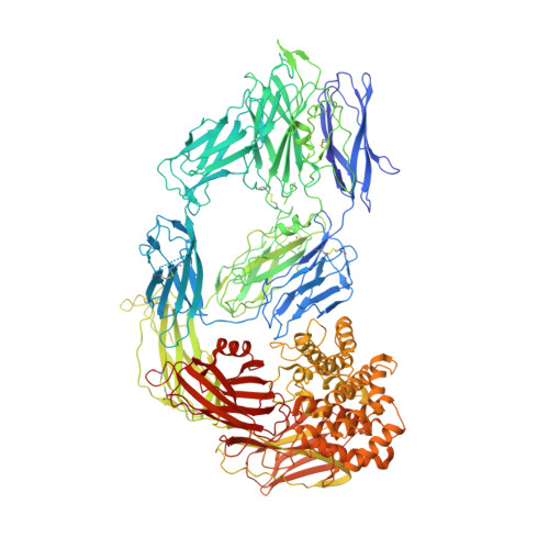
    Alpha-2-Macroglobulin (A2M) is the critical pan-protease inhibitor of the innate immune system. When proteases cleave the A2M bait region, global structural transformation of the A2M tetramer is triggered to entrap the protease. The structural basis behind the cleavage-induced transformation and the protease entrapment remains unclear. Here, we report cryo-EM structures of native- and intermediate-forms of the Xenopus laevis egg A2M homolog (A2Moo or ovomacroglobulin) tetramer at 3.7-4.1 Å and 6.4 Å resolution, respectively. In the native A2Moo tetramer, two pairs of dimers arrange into a cross-like configuration with four 60 Å-wide bait-exposing grooves. Each bait in the native form threads into an aperture formed by three macroglobulin domains (MG2, MG3, MG6). The bait is released from the narrowed aperture in the induced protomer of the intermediate form. We propose that the intact bait region works as a "latch-lock" to block futile A2M transformation until its protease-mediated cleavage.


  • Organizational Affiliation

    Laboratory of Chromosome and Cell Biology, The Rockefeller University, New York, NY 10065, United States. Electronic address: yarimura@rockefeller.edu.


Macromolecules
Find similar proteins by:  (by identity cutoff)  |  3D Structure
Entity ID: 1
MoleculeChains Sequence LengthOrganismDetailsImage
Alpha 2-macroglobulin1,441Xenopus laevisMutation(s): 0 
UniProt
Find proteins for A0A1L8FIE8 (Xenopus laevis)
Explore A0A1L8FIE8 
Go to UniProtKB:  A0A1L8FIE8
Entity Groups  
Sequence Clusters30% Identity50% Identity70% Identity90% Identity95% Identity100% Identity
UniProt GroupA0A1L8FIE8
Sequence Annotations
Expand
  • Reference Sequence
Small Molecules
Ligands 1 Unique
IDChains Name / Formula / InChI Key2D Diagram3D Interactions
NAG
Query on NAG

Download Ideal Coordinates CCD File 
B [auth A]
C [auth A]
D [auth A]
E [auth A]
F [auth A]
B [auth A],
C [auth A],
D [auth A],
E [auth A],
F [auth A],
G [auth A],
H [auth A],
I [auth A],
J [auth A],
K [auth A],
L [auth A],
M [auth A],
N [auth A],
O [auth A],
P [auth A],
Q [auth A],
R [auth A]
2-acetamido-2-deoxy-beta-D-glucopyranose
C8 H15 N O6
OVRNDRQMDRJTHS-FMDGEEDCSA-N
Experimental Data & Validation

Experimental Data

  • Method: ELECTRON MICROSCOPY
  • Resolution: 3.65 Å
  • Aggregation State: PARTICLE 
  • Reconstruction Method: SINGLE PARTICLE 
EM Software:
TaskSoftware PackageVersion
RECONSTRUCTIONcryoSPARC3.2.0
MODEL REFINEMENTPHENIX1.19.2

Structure Validation

View Full Validation Report


Entry History & Funding Information

Deposition Data


Funding OrganizationLocationGrant Number
National Institutes of Health/National Institute of General Medical Sciences (NIH/NIGMS)JapanR35GM132111
Japan Society for the Promotion of Science (JSPS)JapanJSPS Overseas Research Fellowships

Revision History  (Full details and data files)

  • Version 1.0: 2022-01-05
    Type: Initial release
  • Version 1.1: 2022-01-19
    Changes: Database references