Emergence of an Auxin Sensing Domain in Plant-Associated Bacteria.
Gavira, J.A., Rico-Jimenez, M., Ortega, A., Petukhova, N.V., Bug, D.S., Castellvi, A., Porozov, Y.B., Zhulin, I.B., Krell, T., Matilla, M.A.(2023) mBio 14: e0336322-e0336322
- PubMed: 36602305 Search on PubMedSearch on PubMed Central
- DOI: https://doi.org/10.1128/mbio.03363-22
- Primary Citation Related Structures:
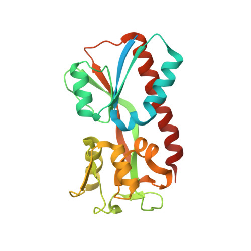
7QEJ, 7QEK - PubMed Abstract:
Bacteria have evolved a sophisticated array of signal transduction systems that allow them to adapt their physiology and metabolism to changing environmental conditions. Typically, these systems recognize signals through dedicated ligand binding domains (LBDs) to ultimately trigger a diversity of physiological responses. Nonetheless, an increasing number of reports reveal that signal transduction receptors also bind antagonists to inhibit responses mediated by agonists. The mechanisms by which antagonists block the downstream signaling cascade remain largely unknown. To advance our knowledge in this field, we used the LysR-type transcriptional regulator AdmX as a model. AdmX activates the expression of an antibiotic biosynthetic cluster in the rhizobacterium Serratia plymuthica. AdmX specifically recognizes the auxin phytohormone indole-3-acetic acid (IAA) and its biosynthetic intermediate indole-3-pyruvic acid (IPA) as signals. However, only IAA, but not IPA, was shown to regulate antibiotic production in S. plymuthica . Here, we report the high-resolution structures of the LBD of AdmX in complex with IAA and IPA. We found that IAA and IPA compete for binding to AdmX. Although IAA and IPA binding does not alter the oligomeric state of AdmX, IPA binding causes a higher degree of compactness in the protein structure. Molecular dynamics simulations revealed significant differences in the binding modes of IAA and IPA by AdmX, and the inspection of the three-dimensional structures evidenced differential agonist- and antagonist-mediated structural changes. Key residues for auxin binding were identified and an auxin recognition motif defined. Phylogenetic clustering supports the recent evolutionary emergence of this motif specifically in plant-associated enterobacteria. IMPORTANCE Although antagonists were found to bind different bacterial signal transduction receptors, we are still at the early stages of understanding the molecular details by which these molecules exert their inhibitory effects. Here, we provide insight into the structural changes resulting from the binding of an agonist and an antagonist to a sensor protein. Our data indicate that agonist and antagonist recognition is characterized by small conformational differences in the LBDs that can be efficiently transmitted to the output domain to modulate the final response. LBDs are subject to strong selective pressures and are rapidly evolving domains. An increasing number of reports support the idea that environmental factors drive the evolution of sensor domains. Given the recent evolutionary history of AdmX homologs, as well as their narrow phyletic distribution within plant-associated bacteria, our results are in accordance with a plant-mediated evolutionary process that resulted in the emergence of receptor proteins that specifically sense auxin phytohormones.
- Laboratory of Crystallographic Studies, IACT (CSIC-UGR), Armilla, Spain.
Organizational Affiliation: