7Q5V

HIF PROLYL HYDROXYLASE 2 (PHD2/EGLN1) IN COMPLEX WITH N-OXALYLGLYCINE (NOG) AND HIF-2 ALPHA CODD (523-542)


Experimental Data Snapshot

  • Method: X-RAY DIFFRACTION
  • Resolution: 1.17 Å
  • R-Value Free: 0.179 
  • R-Value Work: 0.157 
  • R-Value Observed: 0.158 

wwPDB Validation   3D Report Full Report


Ligand Structure Quality Assessment 


This is version 1.3 of the entry. See complete history


Literature

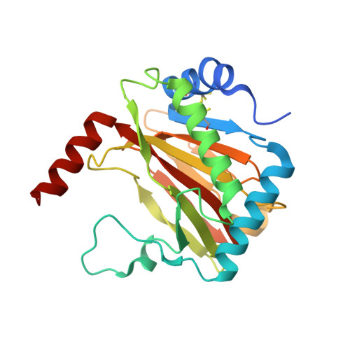
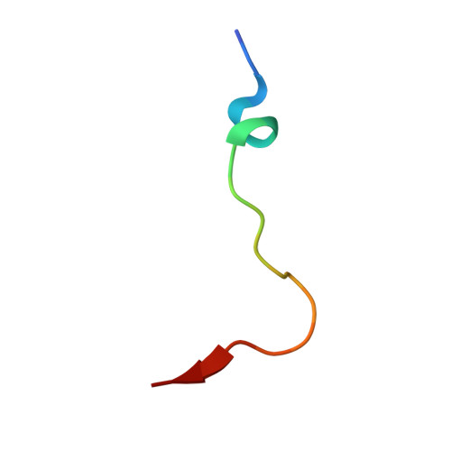
Structural basis for binding of the renal carcinoma target hypoxia-inducible factor 2 alpha to prolyl hydroxylase domain 2.

Figg Jr., W.D.Fiorini, G.Chowdhury, R.Nakashima, Y.Tumber, A.McDonough, M.A.Schofield, C.J.

(2023) Proteins 91: 1510-1524

  • DOI: https://doi.org/10.1002/prot.26541
  • Primary Citation of Related Structures:  
    7Q5V, 7Q5X

  • PubMed Abstract: 

    The hypoxia-inducible factor (HIF) prolyl-hydroxylases (human PHD1-3) catalyze prolyl hydroxylation in oxygen-dependent degradation (ODD) domains of HIFα isoforms, modifications that signal for HIFα proteasomal degradation in an oxygen-dependent manner. PHD inhibitors are used for treatment of anemia in kidney disease. Increased erythropoietin (EPO) in patients with familial/idiopathic erythrocytosis and pulmonary hypertension is associated with mutations in EGLN1 (PHD2) and EPAS1 (HIF2α); a drug inhibiting HIF2α activity is used for clear cell renal cell carcinoma (ccRCC) treatment. We report crystal structures of PHD2 complexed with the C-terminal HIF2α-ODD in the presence of its 2-oxoglutarate cosubstrate or N-oxalylglycine inhibitor. Combined with the reported PHD2.HIFα-ODD structures and biochemical studies, the results inform on the different PHD.HIFα-ODD binding modes and the potential effects of clinically observed mutations in HIFα and PHD2 genes. They may help enable new therapeutic avenues, including PHD isoform-selective inhibitors and sequestration of HIF2α by the PHDs for ccRCC treatment.


  • Organizational Affiliation

    Chemistry Research Laboratory, Department of Chemistry and the Ineos Oxford, Institute for Antimicrobial Research, University of Oxford, Oxford, UK.


Macromolecules
Find similar proteins by:  (by identity cutoff)  |  3D Structure
Entity ID: 1
MoleculeChains Sequence LengthOrganismDetailsImage
Egl nine homolog 1233Homo sapiensMutation(s): 0 
Gene Names: EGLN1C1orf12PNAS-118PNAS-137
EC: 1.14.11.29
UniProt & NIH Common Fund Data Resources
Find proteins for Q9GZT9 (Homo sapiens)
Explore Q9GZT9 
Go to UniProtKB:  Q9GZT9
PHAROS:  Q9GZT9
GTEx:  ENSG00000135766 
Entity Groups  
Sequence Clusters30% Identity50% Identity70% Identity90% Identity95% Identity100% Identity
UniProt GroupQ9GZT9
Sequence Annotations
Expand
  • Reference Sequence

Find similar proteins by:  Sequence   |   3D Structure  

Entity ID: 2
MoleculeChains Sequence LengthOrganismDetailsImage
Endothelial PAS domain-containing protein 120Homo sapiensMutation(s): 0 
UniProt & NIH Common Fund Data Resources
Find proteins for Q99814 (Homo sapiens)
Explore Q99814 
Go to UniProtKB:  Q99814
PHAROS:  Q99814
GTEx:  ENSG00000116016 
Entity Groups  
Sequence Clusters30% Identity50% Identity70% Identity90% Identity95% Identity100% Identity
UniProt GroupQ99814
Sequence Annotations
Expand
  • Reference Sequence
Small Molecules
Ligands 7 Unique
IDChains Name / Formula / InChI Key2D Diagram3D Interactions
OGA (Subject of Investigation/LOI)
Query on OGA

Download Ideal Coordinates CCD File 
D [auth A]N-OXALYLGLYCINE
C4 H5 N O5
BIMZLRFONYSTPT-UHFFFAOYSA-N
PEG
Query on PEG

Download Ideal Coordinates CCD File 
E [auth A],
I [auth A],
J [auth A]
DI(HYDROXYETHYL)ETHER
C4 H10 O3
MTHSVFCYNBDYFN-UHFFFAOYSA-N
GOL
Query on GOL

Download Ideal Coordinates CCD File 
F [auth A]GLYCEROL
C3 H8 O3
PEDCQBHIVMGVHV-UHFFFAOYSA-N
MN
Query on MN

Download Ideal Coordinates CCD File 
C [auth A]MANGANESE (II) ION
Mn
WAEMQWOKJMHJLA-UHFFFAOYSA-N
FMT
Query on FMT

Download Ideal Coordinates CCD File 
G [auth A],
H [auth A],
N [auth B]
FORMIC ACID
C H2 O2
BDAGIHXWWSANSR-UHFFFAOYSA-N
CL
Query on CL

Download Ideal Coordinates CCD File 
K [auth A]CHLORIDE ION
Cl
VEXZGXHMUGYJMC-UHFFFAOYSA-M
MG
Query on MG

Download Ideal Coordinates CCD File 
L [auth A],
M [auth A],
O [auth B]
MAGNESIUM ION
Mg
JLVVSXFLKOJNIY-UHFFFAOYSA-N
Experimental Data & Validation

Experimental Data

  • Method: X-RAY DIFFRACTION
  • Resolution: 1.17 Å
  • R-Value Free: 0.179 
  • R-Value Work: 0.157 
  • R-Value Observed: 0.158 
  • Space Group: P 21 21 2
Unit Cell:
Length ( Å )Angle ( ˚ )
a = 130.914α = 90
b = 38.322β = 90
c = 42.88γ = 90
Software Package:
Software NamePurpose
PHENIXrefinement
xia2data reduction
DIALSdata scaling
PHASERphasing
Cootmodel building
MolProbitymodel building
PDB_EXTRACTdata extraction

Structure Validation

View Full Validation Report


Ligand Structure Quality Assessment 


Entry History & Funding Information

Deposition Data


Funding OrganizationLocationGrant Number
Wellcome TrustUnited Kingdom106244/Z/14/Z
Cancer Research UKUnited Kingdom--

Revision History  (Full details and data files)

  • Version 1.0: 2022-11-16
    Type: Initial release
  • Version 1.1: 2023-07-26
    Changes: Database references
  • Version 1.2: 2023-10-18
    Changes: Data collection, Database references
  • Version 1.3: 2024-01-31
    Changes: Refinement description