7PT1

Actinobacterial 2-hydroxyacyl-CoA lyase (AcHACL) structure in complex with substrate 2-HIB-CoA and inactive cofactor 3-deaza-ThDP


Experimental Data Snapshot

  • Method: X-RAY DIFFRACTION
  • Resolution: 1.55 Å
  • R-Value Free: 0.194 
  • R-Value Work: 0.160 

wwPDB Validation   3D Report Full Report


Ligand Structure Quality Assessment 


This is version 1.2 of the entry. See complete history


Literature

Mechanistic details of the actinobacterial lyase-catalyzed degradation reaction of 2-hydroxyisobutyryl-CoA.

Zahn, M.Konig, G.Pham, H.V.C.Seroka, B.Lazny, R.Yang, G.Ouerfelli, O.Lotowski, Z.Rohwerder, T.

(2022) J Biol Chem 298: 101522-101522

  • DOI: https://doi.org/10.1016/j.jbc.2021.101522
  • Primary Citation of Related Structures:  
    7PT1, 7PT2, 7PT3, 7PT4

  • PubMed Abstract: 

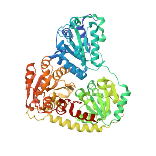
    Actinobacterial 2-hydroxyacyl-CoA lyase reversibly catalyzes the thiamine diphosphate-dependent cleavage of 2-hydroxyisobutyryl-CoA to formyl-CoA and acetone. This enzyme has great potential for use in synthetic one-carbon assimilation pathways for sustainable production of chemicals, but lacks details of substrate binding and reaction mechanism for biochemical reengineering. We determined crystal structures of the tetrameric enzyme in the closed conformation with bound substrate, covalent postcleavage intermediate, and products, shedding light on active site architecture and substrate interactions. Together with molecular dynamics simulations of the covalent precleavage complex, the complete catalytic cycle is structurally portrayed, revealing a proton transfer from the substrate acyl Cβ hydroxyl to residue E493 that returns it subsequently to the postcleavage Cα-carbanion intermediate. Kinetic parameters obtained for mutants E493A, E493Q, and E493K confirm the catalytic role of E493 in the WT enzyme. However, the 10- and 50-fold reduction in lyase activity in the E493A and E493Q mutants, respectively, compared with WT suggests that water molecules may contribute to proton transfer. The putative catalytic glutamate is located on a short α-helix close to the active site. This structural feature appears to be conserved in related lyases, such as human 2-hydroxyacyl-CoA lyase 2. Interestingly, a unique feature of the actinobacterial 2-hydroxyacyl-CoA lyase is a large C-terminal lid domain that, together with active site residues L127 and I492, restricts substrate size to ≤C5 2-hydroxyacyl residues. These details about the catalytic mechanism and determinants of substrate specificity pave the ground for designing tailored catalysts for acyloin condensations for one-carbon and short-chain substrates in biotechnological applications.


  • Organizational Affiliation

    Centre for Enzyme Innovation, School of Biological Sciences, Institute of Biological and Biomedical Sciences, University of Portsmouth, Portsmouth, United Kingdom. Electronic address: michael.zahn@port.ac.uk.


Macromolecules
Find similar proteins by:  (by identity cutoff)  |  3D Structure
Entity ID: 1
MoleculeChains Sequence LengthOrganismDetailsImage
2-hydroxyacyl-CoA lyase
A, B
612Actinomycetospora chiangmaiensis DSM 45062Mutation(s): 0 
EC: 4.2.1.17
UniProt
Find proteins for P0DUV9 (Actinomycetospora chiangmaiensis (strain DSM 45062 / JCM 15998 / CCTCC AA 205017 / NBRC 104400 / YIM 0006))
Explore P0DUV9 
Go to UniProtKB:  P0DUV9
Entity Groups  
Sequence Clusters30% Identity50% Identity70% Identity90% Identity95% Identity100% Identity
UniProt GroupP0DUV9
Sequence Annotations
Expand
  • Reference Sequence
Small Molecules
Ligands 3 Unique
IDChains Name / Formula / InChI Key2D Diagram3D Interactions
3KK (Subject of Investigation/LOI)
Query on 3KK

Download Ideal Coordinates CCD File 
D [auth A],
H [auth B]
S-{(3R,5R,9R)-1-[(2R,3S,4R,5R)-5-(6-amino-9H-purin-9-yl)-4-hydroxy-3-(phosphonooxy)tetrahydrofuran-2-yl]-3,5,9-trihydroxy-8,8-dimethyl-3,5-dioxido-10,14-dioxo-2,4,6-trioxa-11,15-diaza-3lambda~5~,5lambda~5~-diphosphaheptadecan-17-yl} 2-hydroxy-2-methylpropanethioate
C25 H42 N7 O18 P3 S
FFVUICCDNWZCRC-ZSJPKINUSA-N
TPW (Subject of Investigation/LOI)
Query on TPW

Download Ideal Coordinates CCD File 
C [auth A],
G [auth B]
2-{4-[(4-AMINO-2-METHYLPYRIMIDIN-5-YL)METHYL]-3-METHYLTHIOPHEN-2-YL}ETHYL TRIHYDROGEN DIPHOSPHATE
C13 H19 N3 O7 P2 S
IOGGWTLVIZLGGZ-UHFFFAOYSA-N
MG (Subject of Investigation/LOI)
Query on MG

Download Ideal Coordinates CCD File 
E [auth A],
F [auth A],
I [auth B]
MAGNESIUM ION
Mg
JLVVSXFLKOJNIY-UHFFFAOYSA-N
Experimental Data & Validation

Experimental Data

  • Method: X-RAY DIFFRACTION
  • Resolution: 1.55 Å
  • R-Value Free: 0.194 
  • R-Value Work: 0.160 
  • Space Group: C 2 2 21
Unit Cell:
Length ( Å )Angle ( ˚ )
a = 103.95α = 90
b = 146.138β = 90
c = 174.749γ = 90
Software Package:
Software NamePurpose
REFMACrefinement
autoPROCdata reduction
STARANISOdata scaling
MoRDaphasing

Structure Validation

View Full Validation Report


Ligand Structure Quality Assessment 


Entry History & Funding Information

Deposition Data


Funding OrganizationLocationGrant Number
Not funded--

Revision History  (Full details and data files)

  • Version 1.0: 2022-02-02
    Type: Initial release
  • Version 1.1: 2022-02-09
    Changes: Database references
  • Version 1.2: 2024-01-31
    Changes: Data collection, Derived calculations, Refinement description