7MSY

Structure of CalU17 from the Calicheamicin Biosynthesis Pathway of Micromonospora echinospora


Experimental Data Snapshot

  • Method: X-RAY DIFFRACTION
  • Resolution: 2.21 Å
  • R-Value Free: 0.236 
  • R-Value Work: 0.184 
  • R-Value Observed: 0.186 

wwPDB Validation   3D Report Full Report


This is version 1.2 of the entry. See complete history


Literature

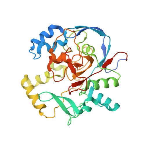
The crystal structure of DynF from the dynemicin-biosynthesis pathway of Micromonospora chersina.

Kosgei, A.J.Miller, M.D.Bhardwaj, M.Xu, W.Thorson, J.S.Van Lanen, S.G.Phillips Jr., G.N.

(2022) Acta Crystallogr F Struct Biol Commun 78: 1-7

  • DOI: https://doi.org/10.1107/S2053230X21012322
  • Primary Citation of Related Structures:  
    6UBL, 7ML6, 7MSY

  • PubMed Abstract: 

    Dynemicin is an enediyne natural product from Micromonospora chersina ATCC53710. Access to the biosynthetic gene cluster of dynemicin has enabled the in vitro study of gene products within the cluster to decipher their roles in assembling this unique molecule. This paper reports the crystal structure of DynF, the gene product of one of the genes within the biosynthetic gene cluster of dynemicin. DynF is revealed to be a dimeric eight-stranded β-barrel structure with palmitic acid bound within a cavity. The presence of palmitic acid suggests that DynF may be involved in binding the precursor polyene heptaene, which is central to the synthesis of the ten-membered ring of the enediyne core.


  • Organizational Affiliation

    Department of Biosciences, Rice University, Houston, TX 77251, USA.


Macromolecules
Find similar proteins by:  (by identity cutoff)  |  3D Structure
Entity ID: 1
MoleculeChains Sequence LengthOrganismDetailsImage
CalU17317Micromonospora echinosporaMutation(s): 0 
Gene Names: calU17
UniProt
Find proteins for Q8KND1 (Micromonospora echinospora)
Explore Q8KND1 
Go to UniProtKB:  Q8KND1
Entity Groups  
Sequence Clusters30% Identity50% Identity70% Identity90% Identity95% Identity100% Identity
UniProt GroupQ8KND1
Sequence Annotations
Expand
  • Reference Sequence
Small Molecules
Ligands 4 Unique
IDChains Name / Formula / InChI Key2D Diagram3D Interactions
GOL
Query on GOL

Download Ideal Coordinates CCD File 
C [auth A]
D [auth A]
E [auth A]
F [auth A]
G [auth A]
C [auth A],
D [auth A],
E [auth A],
F [auth A],
G [auth A],
H [auth A]
GLYCEROL
C3 H8 O3
PEDCQBHIVMGVHV-UHFFFAOYSA-N
CA
Query on CA

Download Ideal Coordinates CCD File 
B [auth A]CALCIUM ION
Ca
BHPQYMZQTOCNFJ-UHFFFAOYSA-N
CL
Query on CL

Download Ideal Coordinates CCD File 
J [auth A],
K [auth A]
CHLORIDE ION
Cl
VEXZGXHMUGYJMC-UHFFFAOYSA-M
MG
Query on MG

Download Ideal Coordinates CCD File 
I [auth A]MAGNESIUM ION
Mg
JLVVSXFLKOJNIY-UHFFFAOYSA-N
Experimental Data & Validation

Experimental Data

  • Method: X-RAY DIFFRACTION
  • Resolution: 2.21 Å
  • R-Value Free: 0.236 
  • R-Value Work: 0.184 
  • R-Value Observed: 0.186 
  • Space Group: P 43 21 2
Unit Cell:
Length ( Å )Angle ( ˚ )
a = 53.465α = 90
b = 53.465β = 90
c = 223.468γ = 90
Software Package:
Software NamePurpose
PHENIXrefinement
PHENIXphasing
XSCALEdata scaling
XDSdata reduction
PDB_EXTRACTdata extraction

Structure Validation

View Full Validation Report


Entry History & Funding Information

Deposition Data


Funding OrganizationLocationGrant Number
National Institutes of Health/National Institute of General Medical Sciences (NIH/NIGMS)United StatesR01-GM115261
National Institutes of Health/National Cancer Institute (NIH/NCI)United StatesR01-CA217255
National Science Foundation (NSF, United States)United StatesSTC-1231306

Revision History  (Full details and data files)

  • Version 1.0: 2021-07-28
    Type: Initial release
  • Version 1.1: 2022-03-02
    Changes: Database references
  • Version 1.2: 2023-10-18
    Changes: Data collection, Refinement description