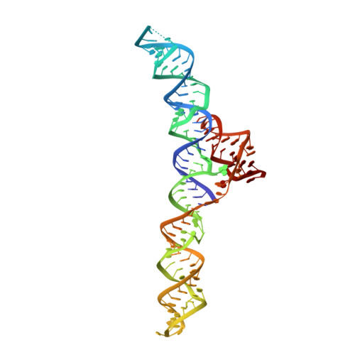
7MLW

Burkholderia sp. TJI49 Guanidine-I riboswitch


Experimental Data Snapshot

  • Method: X-RAY DIFFRACTION
  • Resolution: 2.70 Å
  • R-Value Free: 0.270 
  • R-Value Work: 0.218 
  • R-Value Observed: 0.223 

wwPDB Validation   3D Report Full Report


Ligand Structure Quality Assessment 


This is version 1.1 of the entry. See complete history


Literature

An uncommon [K + (Mg 2+ ) 2 ] metal ion triad imparts stability and selectivity to the Guanidine-I riboswitch.

Trachman 3rd, R.J.Ferre-D'Amare, A.R.

(2021) RNA 27: 1257-1264

  • DOI: https://doi.org/10.1261/rna.078824.121
  • Primary Citation of Related Structures:  
    7MLW

  • PubMed Abstract: 

    The widespread ykkC -I riboswitch class exemplifies divergent riboswitch evolution. To analyze how natural selection has diversified its versatile RNA fold, we determined the X-ray crystal structure of the Burkholderia sp. TJI49 ykkC -I subtype-1 (Guanidine-I) riboswitch aptamer domain. Differing from the previously reported structures of orthologs from Dickeya dadantii and Sulfobacillus acidophilus , our Burkholderia structure reveals a chelated K + ion adjacent to two Mg 2+ ions in the guanidine-binding pocket. Thermal melting analysis shows that K + chelation, which induces localized conformational changes in the binding pocket, improves guanidinium-RNA interactions. Analysis of ribosome structures suggests that the [K + (Mg 2+ ) 2 ] ion triad is uncommon. It is, however, reminiscent of metal ion clusters found in the active sites of ribozymes and DNA polymerases. Previous structural characterization of ykkC -I subtype-2 RNAs, which bind the effector ligands ppGpp and PRPP, indicate that in those paralogs, an adenine responsible for K + chelation in the Burkholderia Guanidine-I riboswitch is replaced by a pyrimidine. This mutation results in a water molecule and Mg 2+ ion binding in place of the K + ion. Thus, our structural analysis demonstrates how ion and solvent chelation tune divergent ligand specificity and affinity among ykkC -I riboswitches.


  • Organizational Affiliation

    Biochemistry and Biophysics Center, National Heart, Lung, and Blood Institute, Bethesda, Maryland 20892-8012, USA.


Macromolecules
Find similar nucleic acids by:  (by identity cutoff)  |  3D Structure
Entity ID: 1
MoleculeChains LengthOrganismImage
Guanidine-I riboswitchA [auth F]128Burkholderia sp. TJI49
Sequence Annotations
Expand
  • Reference Sequence
Small Molecules
Ligands 5 Unique
IDChains Name / Formula / InChI Key2D Diagram3D Interactions
PO4
Query on PO4

Download Ideal Coordinates CCD File 
C [auth F],
D [auth F],
E [auth F],
F
PHOSPHATE ION
O4 P
NBIIXXVUZAFLBC-UHFFFAOYSA-K
SR
Query on SR

Download Ideal Coordinates CCD File 
G [auth F],
H [auth F]
STRONTIUM ION
Sr
PWYYWQHXAPXYMF-UHFFFAOYSA-N
GAI (Subject of Investigation/LOI)
Query on GAI

Download Ideal Coordinates CCD File 
B [auth F]GUANIDINE
C H5 N3
ZRALSGWEFCBTJO-UHFFFAOYSA-N
K (Subject of Investigation/LOI)
Query on K

Download Ideal Coordinates CCD File 
L [auth F],
M [auth F],
N [auth F]
POTASSIUM ION
K
NPYPAHLBTDXSSS-UHFFFAOYSA-N
MG (Subject of Investigation/LOI)
Query on MG

Download Ideal Coordinates CCD File 
I [auth F],
J [auth F],
K [auth F]
MAGNESIUM ION
Mg
JLVVSXFLKOJNIY-UHFFFAOYSA-N
Experimental Data & Validation

Experimental Data

  • Method: X-RAY DIFFRACTION
  • Resolution: 2.70 Å
  • R-Value Free: 0.270 
  • R-Value Work: 0.218 
  • R-Value Observed: 0.223 
  • Space Group: P 32 2 1
Unit Cell:
Length ( Å )Angle ( ˚ )
a = 51.703α = 90
b = 51.703β = 90
c = 266.728γ = 120
Software Package:
Software NamePurpose
PHENIXrefinement
XDSdata reduction
XDSdata scaling
PHASERphasing

Structure Validation

View Full Validation Report


Ligand Structure Quality Assessment 


Entry History 

Deposition Data

Revision History  (Full details and data files)

  • Version 1.0: 2022-01-12
    Type: Initial release
  • Version 1.1: 2023-10-18
    Changes: Data collection, Refinement description