7LZA | pdb_00007lza

Activated form of VanR from S. coelicolor


Experimental Data Snapshot

  • Method: X-RAY DIFFRACTION
  • Resolution: 2.03 Å
  • R-Value Free: 
    0.222 (Depositor), 0.222 (DCC) 
  • R-Value Work: 
    0.182 (Depositor), 0.182 (DCC) 

Starting Models: experimental
View more details

wwPDB Validation 3D Report Full Report

Validation slider image for 7LZA

This is version 1.2 of the entry. See complete history

Literature

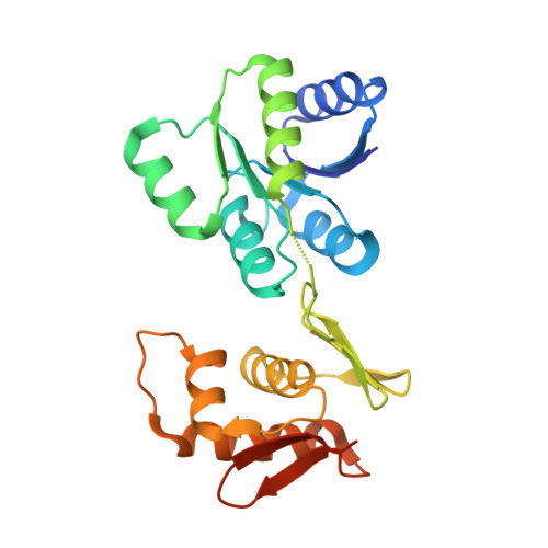
Structures of full-length VanR from Streptomyces coelicolor in both the inactive and activated states.

Maciunas, L.J.Porter, N.Lee, P.J.Gupta, K.Loll, P.J.

(2021) Acta Crystallogr D Struct Biol 77: 1027-1039

  • DOI: https://doi.org/10.1107/S2059798321006288
  • Primary Citation Related Structures: 
    7LZ9, 7LZA

  • PubMed Abstract: 

    Vancomycin has historically been used as a last-resort treatment for serious bacterial infections. However, vancomycin resistance has become widespread in certain pathogens, presenting a serious threat to public health. Resistance to vancomycin is conferred by a suite of resistance genes, the expression of which is controlled by the VanR-VanS two-component system. VanR is the response regulator in this system; in the presence of vancomycin, VanR accepts a phosphoryl group from VanS, thereby activating VanR as a transcription factor and inducing expression of the resistance genes. This paper presents the X-ray crystal structures of full-length VanR from Streptomyces coelicolor in both the inactive and activated states at resolutions of 2.3 and 2.0 Å, respectively. Comparison of the two structures illustrates that phosphorylation of VanR is accompanied by a disorder-to-order transition of helix 4, which lies within the receiver domain of the protein. This transition generates an interface that promotes dimerization of the receiver domain; dimerization in solution was verified using analytical ultracentrifugation. The inactive conformation of the protein does not appear intrinsically unable to bind DNA; rather, it is proposed that in the activated form DNA binding is enhanced by an avidity effect contributed by the receiver-domain dimerization.


  • Organizational Affiliation
    • Department of Biochemistry and Molecular Biology, Drexel University College of Medicine, Philadelphia, PA 19102, USA.

Macromolecule Content 

  • Total Structure Weight: 25.07 kDa 
  • Atom Count: 1,874 
  • Modeled Residue Count: 219 
  • Deposited Residue Count: 232 
  • Unique protein chains: 1

Macromolecules

Find similar proteins by:|  3D Structure
Entity ID: 1
MoleculeChains  Sequence LengthOrganismDetailsImage
Putative two-component system response regulator232Streptomyces coelicolor A3(2)Mutation(s): 0 
Gene Names: SCO3590
UniProt
Find proteins for Q8CJW1 (Streptomyces coelicolor (strain ATCC BAA-471 / A3(2) / M145))
Explore Q8CJW1 
Go to UniProtKB:  Q8CJW1
Entity Groups
Sequence Clusters30% Identity50% Identity70% Identity90% Identity95% Identity100% Identity
UniProt GroupQ8CJW1
Sequence Annotations
Expand
Reference Sequence

Experimental Data & Validation

Experimental Data

  • Method: X-RAY DIFFRACTION
  • Resolution: 2.03 Å
  • R-Value Free:  0.222 (Depositor), 0.222 (DCC) 
  • R-Value Work:  0.182 (Depositor), 0.182 (DCC) 
Space Group: P 65 2 2
Unit Cell:
Length ( Å )Angle ( ˚ )
a = 97.37α = 90
b = 97.37β = 90
c = 118.65γ = 120
Software Package:
Software NamePurpose
XDSdata reduction
XSCALEdata scaling
PHASERphasing
PHENIXrefinement
PDB_EXTRACTdata extraction

Structure Validation

View Full Validation Report


Entry History 

& Funding Information

Deposition Data


Funding OrganizationLocationGrant Number
National Institutes of Health/National Institute Of Allergy and Infectious Diseases (NIH/NIAID)United StatesR01 AI 148679
National Institutes of Health/National Institute Of Allergy and Infectious Diseases (NIH/NIAID)United StatesF31 AI 136385

Revision History  (Full details and data files)

  • Version 1.0: 2021-07-14
    Type: Initial release
  • Version 1.1: 2021-08-25
    Changes: Database references
  • Version 1.2: 2023-10-18
    Changes: Data collection, Refinement description