7LNQ | pdb_00007lnq

Structure of the avibactam-CDD-1 3 minute complex in imidazole and MPD


Experimental Data Snapshot

  • Method: X-RAY DIFFRACTION
  • Resolution: 1.73 Å
  • R-Value Free: 
    0.213 (Depositor), 0.215 (DCC) 
  • R-Value Work: 
    0.185 (Depositor), 0.186 (DCC) 
  • R-Value Observed: 
    0.186 (Depositor) 

Starting Model: experimental
View more details

wwPDB Validation 3D Report Full Report

Validation slider image for 7LNQ

Ligand Structure Quality Assessment 


This is version 1.3 of the entry. See complete history

Literature

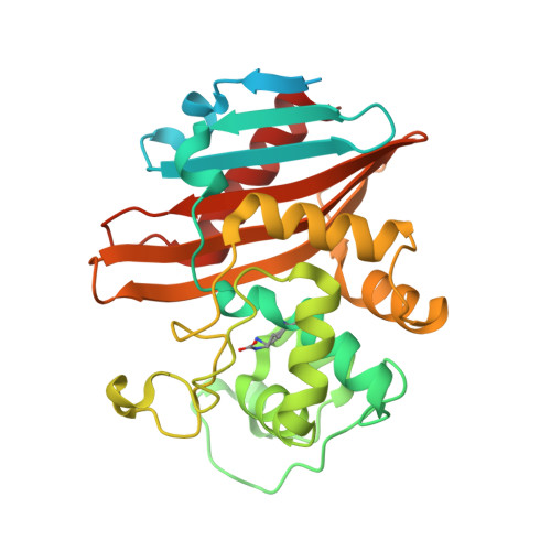
In Crystallo Time-Resolved Interaction of the Clostridioides difficile CDD-1 enzyme with Avibactam Provides New Insights into the Catalytic Mechanism of Class D beta-lactamases.

Stewart, N.K.Toth, M.Stasyuk, A.Vakulenko, S.B.Smith, C.A.

(2021) ACS Infect Dis 7: 1765-1776

  • DOI: https://doi.org/10.1021/acsinfecdis.1c00094
  • Primary Citation Related Structures: 
    7LNO, 7LNQ, 7LNR, 7ME9, 7MEA, 7MEB, 7MEC, 7MED, 7MEE, 7MEF, 7MEG, 7MEH

  • PubMed Abstract: 

    Class D β-lactamases have risen to notoriety due to their wide spread in bacterial pathogens, propensity to inactivate clinically important β-lactam antibiotics, and ability to withstand inhibition by the majority of classical β-lactamase inhibitors. Understanding the catalytic mechanism of these enzymes is thus vitally important for the development of novel antibiotics and inhibitors active against infections caused by antibiotic-resistant bacteria. Here we report an in crystallo time-resolved study of the interaction of the class D β-lactamase CDD-1 from Clostridioides difficile with the diazobicyclooctane inhibitor, avibactam. We show that the catalytic carboxylated lysine, a residue that is essential for both acylation and deacylation of β-lactams, is sequestered within an internal sealed pocket of the enzyme. Time-resolved snapshots generated in this study allowed us to observe decarboxylation of the lysine and movement of CO 2 and water molecules through a transient channel formed between the lysine pocket and the substrate binding site facilitated by rotation of the side chain of a conserved leucine residue. These studies provide novel insights on avibactam binding to CDD-1 and into the catalytic mechanism of class D β-lactamases in general.


  • Organizational Affiliation
    • Department of Chemistry and Biochemistry, University of Notre Dame, Notre Dame, Indiana 46556, United States.

Macromolecule Content 

  • Total Structure Weight: 36.86 kDa 
  • Atom Count: 2,302 
  • Modeled Residue Count: 251 
  • Deposited Residue Count: 312 
  • Unique protein chains: 1

Macromolecules

Find similar proteins by:|  3D Structure
Entity ID: 1
MoleculeChains  Sequence LengthOrganismDetailsImage
Beta-lactamase312Clostridioides difficileMutation(s): 2 
Gene Names: blaR1_4blaR1_1E5F39_11445SAMEA2239407_03320SAMEA3374989_01677
EC: 3.5.2.6

Small Molecules

Ligands 4 Unique
IDChains Name / Formula / InChI Key2D Diagram3D Interactions
NXL
(Subject of Investigation/LOI)

Query on NXL


Download:Ideal Coordinates CCD File
B [auth A](2S,5R)-1-formyl-5-[(sulfooxy)amino]piperidine-2-carboxamide
C7 H13 N3 O6 S
WJDGWXPPFHLLNL-RITPCOANSA-N
MPD

Query on MPD


Download:Ideal Coordinates CCD File
H [auth A](4S)-2-METHYL-2,4-PENTANEDIOL
C6 H14 O2
SVTBMSDMJJWYQN-YFKPBYRVSA-N
SO4

Query on SO4


Download:Ideal Coordinates CCD File
D [auth A],
E [auth A],
F [auth A],
G [auth A]
SULFATE ION
O4 S
QAOWNCQODCNURD-UHFFFAOYSA-L
CO2

Query on CO2


Download:Ideal Coordinates CCD File
C [auth A]CARBON DIOXIDE
C O2
CURLTUGMZLYLDI-UHFFFAOYSA-N
Modified Residues  1 Unique
IDChains TypeFormula2D DiagramParent
KCX
Query on KCX
A
L-PEPTIDE LINKINGC7 H14 N2 O4LYS

Experimental Data & Validation

Experimental Data

  • Method: X-RAY DIFFRACTION
  • Resolution: 1.73 Å
  • R-Value Free:  0.213 (Depositor), 0.215 (DCC) 
  • R-Value Work:  0.185 (Depositor), 0.186 (DCC) 
  • R-Value Observed: 0.186 (Depositor) 
Space Group: P 43 3 2
Unit Cell:
Length ( Å )Angle ( ˚ )
a = 123.548α = 90
b = 123.548β = 90
c = 123.548γ = 90
Software Package:
Software NamePurpose
PHENIXrefinement
PDB_EXTRACTdata extraction
XDSdata reduction
Aimlessdata scaling
REFMACrefinement

Structure Validation

View Full Validation Report


Ligand Structure Quality Assessment 


Entry History 

Deposition Data

Revision History  (Full details and data files)

  • Version 1.0: 2021-05-12
    Type: Initial release
  • Version 1.1: 2021-06-23
    Changes: Database references
  • Version 1.2: 2023-10-18
    Changes: Data collection, Database references, Refinement description
  • Version 1.3: 2023-11-15
    Changes: Data collection