Structural and biochemical analysis of human ADP-ribosyl-acceptor hydrolase 3 reveals the basis of metal selectivity and different roles for the two magnesium ions.
Pourfarjam, Y., Ma, Z., Kurinov, I., Moss, J., Kim, I.K.(2021) J Biol Chem 296: 100692-100692
- PubMed: 33894202 Search on PubMedSearch on PubMed Central
- DOI: https://doi.org/10.1016/j.jbc.2021.100692
- Primary Citation Related Structures:
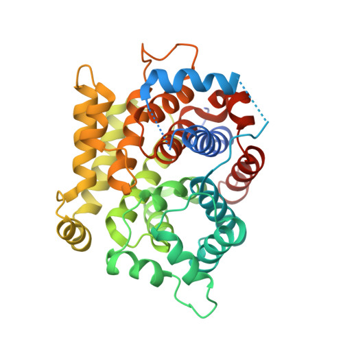
7L9F, 7L9H, 7L9I - PubMed Abstract:
ADP-ribosylation is a reversible and site-specific post-translational modification that regulates a wide array of cellular signaling pathways. Regulation of ADP-ribosylation is vital for maintaining genomic integrity, and uncontrolled accumulation of poly(ADP-ribosyl)ation triggers a poly(ADP-ribose) (PAR)-dependent release of apoptosis-inducing factor from mitochondria, leading to cell death. ADP-ribosyl-acceptor hydrolase 3 (ARH3) cleaves PAR and mono(ADP-ribosyl)ation at serine following DNA damage. ARH3 is also a metalloenzyme with strong metal selectivity. While coordination of two magnesium ions (Mg A and Mg B ) significantly enhances its catalytic efficiency, calcium binding suppresses its function. However, how the coordination of different metal ions affects its catalysis has not been defined. Here, we report a new crystal structure of ARH3 complexed with its product ADP-ribose and calcium. This structure shows that calcium coordination significantly distorts the binuclear metal center of ARH3, which results in decreased binding affinity to ADP-ribose, and suboptimal substrate alignment, leading to impaired hydrolysis of PAR and mono(ADP-ribosyl)ated serines. Furthermore, combined structural and mutational analysis of the metal-coordinating acidic residues revealed that Mg A is crucial for optimal substrate positioning for catalysis, whereas Mg B plays a key role in substrate binding. Our collective data provide novel insights into the different roles of these metal ions and the basis of metal selectivity of ARH3 and contribute to understanding the dynamic regulation of cellular ADP-ribosylations during the DNA damage response.
- Department of Chemistry, University of Cincinnati, Cincinnati, Ohio, USA.
Organizational Affiliation: