7KDV

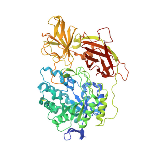
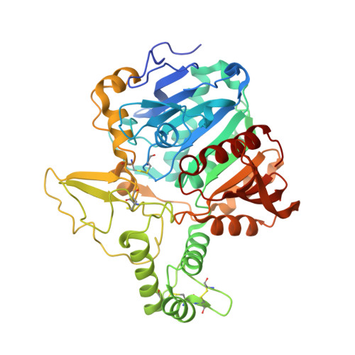
Murine core lysosomal multienzyme complex (LMC) composed of acid beta-galactosidase (GLB1) and protective protein cathepsin A (PPCA, CTSA)


Experimental Data Snapshot

  • Method: ELECTRON MICROSCOPY
  • Resolution: 4.59 Å
  • Aggregation State: PARTICLE 
  • Reconstruction Method: SINGLE PARTICLE 

wwPDB Validation   3D Report Full Report


This is version 1.1 of the entry. See complete history


Literature

Structure of the murine lysosomal multienzyme complex core.

Gorelik, A.Illes, K.Hasan, S.M.N.Nagar, B.Mazhab-Jafari, M.T.

(2021) Sci Adv 7

  • DOI: https://doi.org/10.1126/sciadv.abf4155
  • Primary Citation of Related Structures:  
    7KDV

  • PubMed Abstract: 

    The enzymes β-galactosidase (GLB1) and neuraminidase 1 (NEU1; sialidase 1) participate in the degradation of glycoproteins and glycolipids in the lysosome. To remain active and stable, they associate with PPCA [protective protein cathepsin A (CTSA)] into a high-molecular weight lysosomal multienzyme complex (LMC), of which several forms exist. Genetic defects in these three proteins cause the lysosomal storage diseases GM1-gangliosidosis/mucopolysaccharidosis IV type B, sialidosis, and galactosialidosis, respectively. To better understand the interactions between these enzymes, we determined the three-dimensional structure of the murine LMC core. This 0.8-MDa complex is composed of three GLB1 dimers and three CTSA dimers, adopting a triangular architecture maintained through six copies of a unique GLB1-CTSA polar interface. Mutations in this contact surface that occur in GM1-gangliosidosis prevent formation of the LMC in vitro. These findings may facilitate development of therapies for lysosomal storage disorders.


  • Organizational Affiliation

    Department of Biochemistry, McGill University, Montreal, Quebec, Canada.


Macromolecules
Find similar proteins by:  (by identity cutoff)  |  3D Structure
Entity ID: 1
MoleculeChains Sequence LengthOrganismDetailsImage
Beta-galactosidase
A, C, E, G, I
A, C, E, G, I, K
630Mus musculusMutation(s): 3 
Gene Names: Glb1BglGlb-1
EC: 3.2.1.23
UniProt
Find proteins for P23780 (Mus musculus)
Explore P23780 
Go to UniProtKB:  P23780
Entity Groups  
Sequence Clusters30% Identity50% Identity70% Identity90% Identity95% Identity100% Identity
UniProt GroupP23780
Sequence Annotations
Expand
  • Reference Sequence
Find similar proteins by:  (by identity cutoff)  |  3D Structure
Entity ID: 2
MoleculeChains Sequence LengthOrganismDetailsImage
Lysosomal protective protein
B, D, F, H, J
B, D, F, H, J, L
461Mus musculusMutation(s): 1 
Gene Names: CtsaPpgb
EC: 3.4.16.5
UniProt & NIH Common Fund Data Resources
Find proteins for P16675 (Mus musculus)
Explore P16675 
Go to UniProtKB:  P16675
IMPC:  MGI:97748
Entity Groups  
Sequence Clusters30% Identity50% Identity70% Identity90% Identity95% Identity100% Identity
UniProt GroupP16675
Sequence Annotations
Expand
  • Reference Sequence
Oligosaccharides

Help

Entity ID: 3
MoleculeChains Length2D Diagram Glycosylation3D Interactions
2-acetamido-2-deoxy-beta-D-glucopyranose-(1-4)-2-acetamido-2-deoxy-beta-D-glucopyranose
BA [auth b],
CA [auth c],
M,
N,
P,
BA [auth b],
CA [auth c],
M,
N,
P,
Q,
S,
T,
V,
W,
Y,
Z
2N-Glycosylation
Glycosylation Resources
GlyTouCan:  G42666HT
GlyCosmos:  G42666HT
GlyGen:  G42666HT
Entity ID: 4
MoleculeChains Length2D Diagram Glycosylation3D Interactions
beta-D-mannopyranose-(1-4)-2-acetamido-2-deoxy-beta-D-glucopyranose-(1-4)-2-acetamido-2-deoxy-beta-D-glucopyranose
AA [auth a],
DA [auth d],
O,
R,
U,
AA [auth a],
DA [auth d],
O,
R,
U,
X
3N-Glycosylation
Glycosylation Resources
GlyTouCan:  G15407YE
GlyCosmos:  G15407YE
GlyGen:  G15407YE
Small Molecules
Ligands 1 Unique
IDChains Name / Formula / InChI Key2D Diagram3D Interactions
NAG
Query on NAG

Download Ideal Coordinates CCD File 
AB [auth I]
BB [auth I]
CB [auth J]
DB [auth K]
EA [auth A]
AB [auth I],
BB [auth I],
CB [auth J],
DB [auth K],
EA [auth A],
EB [auth K],
FA [auth A],
FB [auth K],
GA [auth A],
GB [auth K],
HA [auth A],
HB [auth L],
IA [auth B],
JA [auth C],
KA [auth C],
LA [auth C],
MA [auth C],
NA [auth D],
OA [auth E],
PA [auth E],
QA [auth E],
RA [auth E],
SA [auth F],
TA [auth G],
UA [auth G],
VA [auth G],
WA [auth G],
XA [auth H],
YA [auth I],
ZA [auth I]
2-acetamido-2-deoxy-beta-D-glucopyranose
C8 H15 N O6
OVRNDRQMDRJTHS-FMDGEEDCSA-N
Experimental Data & Validation

Experimental Data

  • Method: ELECTRON MICROSCOPY
  • Resolution: 4.59 Å
  • Aggregation State: PARTICLE 
  • Reconstruction Method: SINGLE PARTICLE 
EM Software:
TaskSoftware PackageVersion
MODEL REFINEMENTPHENIX
RECONSTRUCTIONcryoSPARC

Structure Validation

View Full Validation Report


Entry History & Funding Information

Deposition Data


Funding OrganizationLocationGrant Number
Canadian Institutes of Health Research (CIHR)CanadaMFE-164773
Canadian Institutes of Health Research (CIHR)CanadaMOP-133535
Canadian Institutes of Health Research (CIHR)Canada419240

Revision History  (Full details and data files)

  • Version 1.0: 2021-03-17
    Type: Initial release
  • Version 1.1: 2021-05-26
    Changes: Database references