7JI8

Self-assembly of a 3D DNA crystal lattice (4x6 scramble junction version) containing the J31 immobile Holliday junction with R3 symmetry


Experimental Data Snapshot

  • Method: X-RAY DIFFRACTION
  • Resolution: 2.95 Å
  • R-Value Free: 0.281 
  • R-Value Work: 0.255 
  • R-Value Observed: 0.257 

wwPDB Validation   3D Report Full Report


This is version 1.2 of the entry. See complete history


Literature

The influence of Holliday junction sequence and dynamics on DNA crystal self-assembly.

Simmons, C.R.MacCulloch, T.Krepl, M.Matthies, M.Buchberger, A.Crawford, I.Sponer, J.Sulc, P.Stephanopoulos, N.Yan, H.

(2022) Nat Commun 13: 3112-3112

  • DOI: https://doi.org/10.1038/s41467-022-30779-6
  • Primary Citation of Related Structures:  
    6WQG, 6WR3, 6WR5, 6WR7, 6WR9, 6WRA, 6WRB, 6WRC, 6WRI, 6WRJ, 6WSN, 6WSO, 6WSP, 6WSQ, 6WSR, 6WSS, 6WST, 6WSU, 6WSV, 6WSW, 6WSX, 6WSY, 6WSZ, 6WT0, 6WT1, 6X8B, 6X8C, 6XDV, 6XDW, 6XDX, 6XDY, 6XDZ, 6XEI, 6XEJ, 6XEK, 6XEL, 6XEM, 6XFC, 6XFD, 6XFE, 6XFF, 6XFG, 6XFW, 6XFX, 6XFY, 6XFZ, 6XG0, 6XGJ, 6XGK, 6XGL

  • PubMed Abstract: 

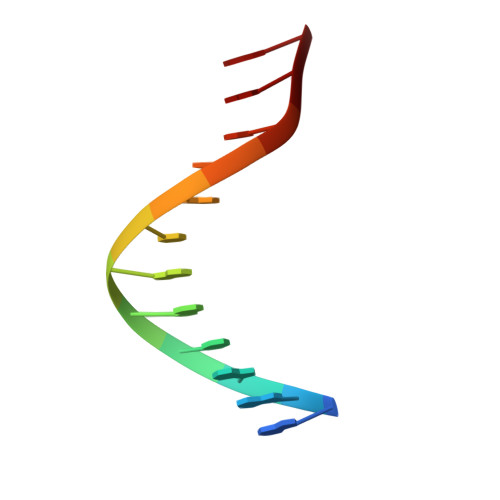
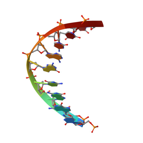
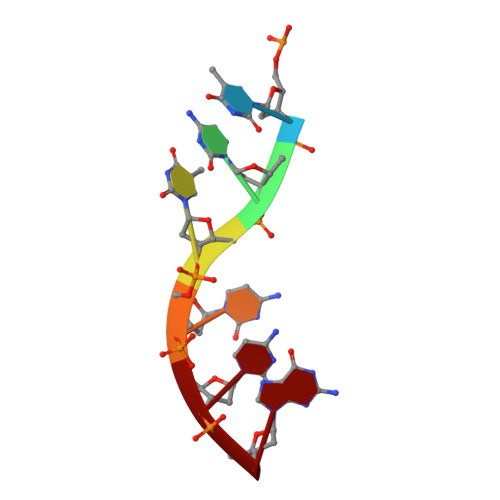
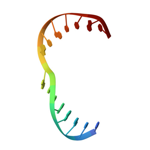
    The programmable synthesis of rationally engineered crystal architectures for the precise arrangement of molecular species is a foundational goal in nanotechnology, and DNA has become one of the most prominent molecules for the construction of these materials. In particular, branched DNA junctions have been used as the central building block for the assembly of 3D lattices. Here, crystallography is used to probe the effect of all 36 immobile Holliday junction sequences on self-assembling DNA crystals. Contrary to the established paradigm in the field, most junctions yield crystals, with some enhancing the resolution or resulting in unique crystal symmetries. Unexpectedly, even the sequence adjacent to the junction has a significant effect on the crystal assemblies. Six of the immobile junction sequences are completely resistant to crystallization and thus deemed "fatal," and molecular dynamics simulations reveal that these junctions invariably lack two discrete ion binding sites that are pivotal for crystal formation. The structures and dynamics detailed here could be used to inform future designs of both crystals and DNA nanostructures more broadly, and have potential implications for the molecular engineering of applied nanoelectronics, nanophotonics, and catalysis within the crystalline context.


  • Organizational Affiliation

    Biodesign Center for Molecular Design and Biomimetics, Arizona State University, 1001S. McAllister Ave, Tempe, AZ, 85287, USA.


Macromolecules

Find similar nucleic acids by:  Sequence   |   3D Structure  

Entity ID: 1
MoleculeChains LengthOrganismImage
DNA (5'-D(*GP*AP*AP*CP*GP*AP*CP*AP*CP*AP*GP*A)-3')12synthetic construct
Sequence Annotations
Expand
  • Reference Sequence

Find similar nucleic acids by:  Sequence   |   3D Structure  

Entity ID: 2
MoleculeChains LengthOrganismImage
DNA (5'-D(P*CP*GP*GP*CP*GP*AP*CP*TP*C)-3')9synthetic construct
Sequence Annotations
Expand
  • Reference Sequence

Find similar nucleic acids by:  Sequence   |   3D Structure  

Entity ID: 3
MoleculeChains LengthOrganismImage
DNA (5'-D(P*TP*CP*TP*CP*CP*G)-3')6synthetic construct
Sequence Annotations
Expand
  • Reference Sequence

Find similar nucleic acids by:  Sequence   |   3D Structure  

Entity ID: 4
MoleculeChains LengthOrganismImage
DNA (5'-D(*TP*CP*GP*AP*GP*TP*CP*GP*GP*TP*GP*TP*CP*GP*T)-3')15synthetic construct
Sequence Annotations
Expand
  • Reference Sequence
Small Molecules
Ligands 1 Unique
IDChains Name / Formula / InChI Key2D Diagram3D Interactions
CAC
Query on CAC

Download Ideal Coordinates CCD File 
E [auth C]CACODYLATE ION
C2 H6 As O2
OGGXGZAMXPVRFZ-UHFFFAOYSA-M
Experimental Data & Validation

Experimental Data

  • Method: X-RAY DIFFRACTION
  • Resolution: 2.95 Å
  • R-Value Free: 0.281 
  • R-Value Work: 0.255 
  • R-Value Observed: 0.257 
  • Space Group: H 3
Unit Cell:
Length ( Å )Angle ( ˚ )
a = 113.109α = 90
b = 113.109β = 90
c = 50.385γ = 120
Software Package:
Software NamePurpose
HKL-2000data reduction
HKL-2000data scaling
PHENIXrefinement
PDB_EXTRACTdata extraction
PHASERphasing

Structure Validation

View Full Validation Report


Entry History & Funding Information

Deposition Data


Funding OrganizationLocationGrant Number
National Science Foundation (NSF, United States)United States1360635
National Institutes of Health/National Institute of General Medical Sciences (NIH/NIGMS)United StatesR01GM104960
National Science Foundation (NSF, United States)United StatesNSF2004250

Revision History  (Full details and data files)

  • Version 1.0: 2021-07-14
    Type: Initial release
  • Version 1.1: 2022-07-06
    Changes: Database references
  • Version 1.2: 2023-10-18
    Changes: Data collection, Refinement description