


Neutron crystallographic analysis of the nucleotide-binding domain of Hsp72 in complex with ADP.
Yokoyama, T., Fujii, S., Ostermann, A., Schrader, T.E., Nabeshima, Y., Mizuguchi, M.(2022) IUCrJ 9: 562-572
Experimental Data Snapshot
Starting Model: experimental
View more details
(2022) IUCrJ 9: 562-572
Entity ID: 1 | |||||
|---|---|---|---|---|---|
| Molecule | Chains | Sequence Length | Organism | Details | Image |
| Heat shock 70 kDa protein 1B | 386 | Homo sapiens | Mutation(s): 1 Gene Names: HSPA1B, HSP72 | 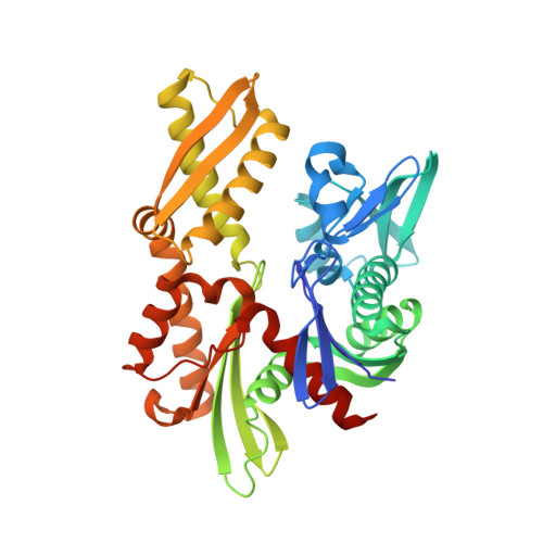 | |
UniProt & NIH Common Fund Data Resources | |||||
PHAROS: P0DMV9 | |||||
Entity Groups | |||||
| Sequence Clusters | 30% Identity50% Identity70% Identity90% Identity95% Identity100% Identity | ||||
| UniProt Group | P0DMV9 | ||||
Sequence AnnotationsExpand | |||||
Reference Sequence | |||||
| Ligands 3 Unique | |||||
|---|---|---|---|---|---|
| ID | Chains | Name / Formula / InChI Key | 2D Diagram | 3D Interactions | |
| ANP (Subject of Investigation/LOI) Download:Ideal Coordinates CCD File | E [auth A] | PHOSPHOAMINOPHOSPHONIC ACID-ADENYLATE ESTER C10 H17 N6 O12 P3 PVKSNHVPLWYQGJ-KQYNXXCUSA-N |  | ||
| CL (Subject of Investigation/LOI) Download:Ideal Coordinates CCD File | D [auth A] | CHLORIDE ION Cl VEXZGXHMUGYJMC-UHFFFAOYSA-M |  | ||
| MG (Subject of Investigation/LOI) Download:Ideal Coordinates CCD File | B [auth A], C [auth A] | MAGNESIUM ION Mg JLVVSXFLKOJNIY-UHFFFAOYSA-N |  | ||
| Length ( Å ) | Angle ( ˚ ) |
|---|---|
| a = 45.827 | α = 90 |
| b = 61.968 | β = 90 |
| c = 142.779 | γ = 90 |
| Software Name | Purpose |
|---|---|
| PHENIX | refinement |
| XDS | data reduction |
| XSCALE | data scaling |
| PHENIX | phasing |


