7EXP | pdb_00007exp

Crystal structure of zebrafish TRAP1 with AMPPNP and MitoQ


Experimental Data Snapshot

  • Method: X-RAY DIFFRACTION
  • Resolution: 2.30 Å
  • R-Value Free: 
    0.241 (Depositor), 0.228 (DCC) 
  • R-Value Work: 
    0.195 (Depositor) 
  • R-Value Observed: 
    0.197 (Depositor) 

Starting Model: experimental
View more details

wwPDB Validation 3D Report Full Report

Validation slider image for 7EXP

Ligand Structure Quality Assessment 


This is version 1.2 of the entry. See complete history

Literature

Mitoquinone Inactivates Mitochondrial Chaperone TRAP1 by Blocking the Client Binding Site.

Yoon, N.G.Lee, H.Kim, S.Y.Hu, S.Kim, D.Yang, S.Hong, K.B.Lee, J.H.Kang, S.Kim, B.G.Myung, K.Lee, C.Kang, B.H.

(2021) J Am Chem Soc 143: 19684-19696

  • DOI: https://doi.org/10.1021/jacs.1c07099
  • Primary Citation Related Structures: 
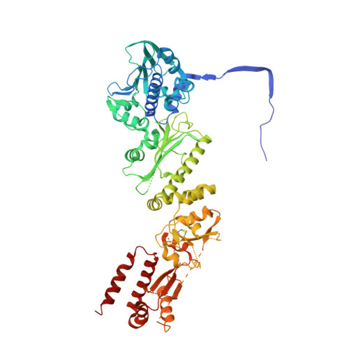
    7EXP

  • PubMed Abstract: 

    Heat shock protein 90 (Hsp90) family proteins are molecular chaperones that modulate the functions of various substrate proteins (clients) implicated in pro-tumorigenic pathways. In this study, the mitochondria-targeted antioxidant mitoquinone (MitoQ) was identified as a potent inhibitor of mitochondrial Hsp90, known as a tumor necrosis factor receptor-associated protein 1 (TRAP1). Structural analyses revealed an asymmetric bipartite interaction between MitoQ and the previously unrecognized drug binding sites located in the middle domain of TRAP1, believed to be a client binding region. MitoQ effectively competed with TRAP1 clients, and MitoQ treatment facilitated the identification of 103 TRAP1-interacting mitochondrial proteins in cancer cells. MitoQ and its redox-crippled SB-U014/SB-U015 exhibited more potent anticancer activity in vitro and in vivo than previously reported mitochondria-targeted TRAP1 inhibitors. The findings indicate that targeting the client binding site of Hsp90 family proteins offers a novel strategy for the development of potent anticancer drugs.


  • Organizational Affiliation
    • Department of Biological Sciences, Ulsan National Institutes of Science and Technology (UNIST), Ulsan 44919, South Korea.

Macromolecule Content 

  • Total Structure Weight: 149.89 kDa 
  • Atom Count: 9,793 
  • Modeled Residue Count: 1,173 
  • Deposited Residue Count: 1,294 
  • Unique protein chains: 1

Macromolecules

Find similar proteins by:|  3D Structure
Entity ID: 1
MoleculeChains  Sequence LengthOrganismDetailsImage
TNF receptor-associated protein 1
A, B
647Danio rerioMutation(s): 0 
Gene Names: trap1
UniProt
Find proteins for A0A2R8RKV0 (Danio rerio)
Explore A0A2R8RKV0 
Go to UniProtKB:  A0A2R8RKV0
Entity Groups
Sequence Clusters30% Identity50% Identity70% Identity90% Identity95% Identity100% Identity
UniProt GroupA0A2R8RKV0
Sequence Annotations
Expand
Reference Sequence

Small Molecules

Ligands 4 Unique
IDChains Name / Formula / InChI Key2D Diagram3D Interactions
05M
(Subject of Investigation/LOI)

Query on 05M


Download:Ideal Coordinates CCD File
G [auth A]2,3-dimethoxy-5-methyl-6-[10-(triphenyl-$l^{5}-phosphanyl)decyl]cyclohexa-2,5-diene-1,4-dione
C37 H45 O4 P
BIPKQWZPPRRUAP-UHFFFAOYSA-N
ANP

Query on ANP


Download:Ideal Coordinates CCD File
C [auth A],
H [auth B]
PHOSPHOAMINOPHOSPHONIC ACID-ADENYLATE ESTER
C10 H17 N6 O12 P3
PVKSNHVPLWYQGJ-KQYNXXCUSA-N
CO

Query on CO


Download:Ideal Coordinates CCD File
E [auth A],
F [auth A],
J [auth B],
K [auth B]
COBALT (II) ION
Co
XLJKHNWPARRRJB-UHFFFAOYSA-N
MG

Query on MG


Download:Ideal Coordinates CCD File
D [auth A],
I [auth B]
MAGNESIUM ION
Mg
JLVVSXFLKOJNIY-UHFFFAOYSA-N

Experimental Data & Validation

Experimental Data

  • Method: X-RAY DIFFRACTION
  • Resolution: 2.30 Å
  • R-Value Free:  0.241 (Depositor), 0.228 (DCC) 
  • R-Value Work:  0.195 (Depositor) 
  • R-Value Observed: 0.197 (Depositor) 
Space Group: C 1 2 1
Unit Cell:
Length ( Å )Angle ( ˚ )
a = 179.339α = 90
b = 96.522β = 134.39
c = 125.615γ = 90
Software Package:
Software NamePurpose
PHENIXrefinement
PDB_EXTRACTdata extraction
PHENIXphasing
HKL-2000data scaling
HKL-2000data collection

Structure Validation

View Full Validation Report


Ligand Structure Quality Assessment 


Entry History 

Deposition Data

Revision History  (Full details and data files)

  • Version 1.0: 2022-01-05
    Type: Initial release
  • Version 1.1: 2023-11-29
    Changes: Data collection, Refinement description
  • Version 1.2: 2025-09-17
    Changes: Advisory, Derived calculations, Structure summary