7EOX

Protease structure from Euphorbia resinifera


Experimental Data Snapshot

  • Method: X-RAY DIFFRACTION
  • Resolution: 1.70 Å
  • R-Value Free: 0.237 
  • R-Value Work: 0.203 
  • R-Value Observed: 0.204 

wwPDB Validation   3D Report Full Report


Ligand Structure Quality Assessment 


This is version 1.1 of the entry. See complete history


Literature

Sequence analysis and crystal structure of a glycosylated protease from Euphorbia resinifera latex for its proteolytic activity aspect.

Siritapetawee, J.Attarataya, J.Charoenwattanasatien, R.

(2022) Biotechnol Appl Biochem 69: 2580-2591

  • DOI: https://doi.org/10.1002/bab.2307
  • Primary Citation of Related Structures:  
    7EOX

  • PubMed Abstract: 

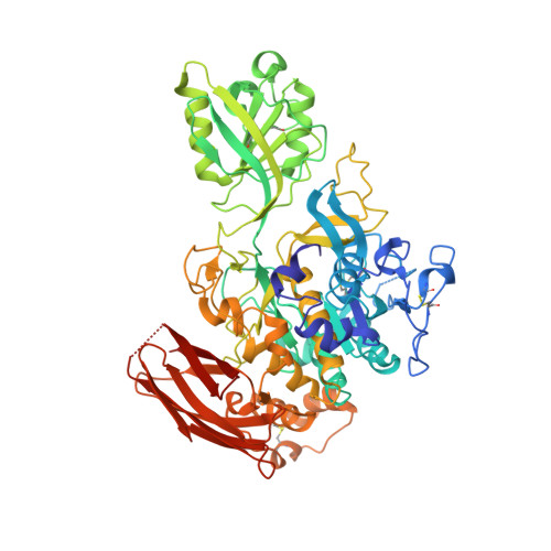
    The investigation of a plant glycosylated serine protease (EuRP-61) isolated from Euphorbia resinifera latex for potential antiplatelet and anticoagulation activities has been previously reported. In the present study, the protein sequence and native crystal structure of EuRP-61 were characterized. The structure was identified using single-wavelength anomalous diffraction with a refinement resolution of 1.7 Å (PDB ID: 7EOX). The main structural components of EuRP-61 were composed of three domains: catalytic, protease-associated (PA), and fibronectin type III (Fn3)-like domains. The crystal structure revealed that some loops in the PA and catalytic domains of EuRP-61 were different from the other subtilisin-like proteases (cucumisin and SBT3). These different loops might be involved in the general monomer formation of EuRP-61, substrate specificity, and maintenance of the catalytic domain. The Fn3-like domain may provide flexibility to the enzyme to bind with various substrates and cell receptors. Additionally, the active site of EuRP-61 consisted of the catalytic triad of Ser434, His106, and Asp32, similar to other serine proteases. The present study provides additional information and insight into the protease and antithrombotic activities of EuRP-61, which could contribute to further development of this enzyme for biomedical treatment.


  • Organizational Affiliation

    Biochemistry-Electrochemistry Research Unit, School of Chemistry, Institute of Science, Suranaree University of Technology, Nakhon Ratchasima, Thailand.


Macromolecules
Find similar proteins by:  (by identity cutoff)  |  3D Structure
Entity ID: 1
MoleculeChains Sequence LengthOrganismDetailsImage
Protease
A, B
654Euphorbia resiniferaMutation(s): 0 
Entity Groups  
Sequence Clusters30% Identity50% Identity70% Identity90% Identity95% Identity100% Identity
Sequence Annotations
Expand
  • Reference Sequence
Oligosaccharides

Help

Entity ID: 2
MoleculeChains Length2D Diagram Glycosylation3D Interactions
beta-D-xylopyranose-(1-2)-[alpha-D-mannopyranose-(1-3)][alpha-D-mannopyranose-(1-6)]beta-D-mannopyranose-(1-4)-2-acetamido-2-deoxy-beta-D-glucopyranose-(1-4)-[alpha-L-fucopyranose-(1-3)]2-acetamido-2-deoxy-beta-D-glucopyranose
C
7N-Glycosylation
Glycosylation Resources
GlyTouCan:  G26295XE
GlyCosmos:  G26295XE
GlyGen:  G26295XE
Experimental Data & Validation

Experimental Data

  • Method: X-RAY DIFFRACTION
  • Resolution: 1.70 Å
  • R-Value Free: 0.237 
  • R-Value Work: 0.203 
  • R-Value Observed: 0.204 
  • Space Group: P 21 21 21
Unit Cell:
Length ( Å )Angle ( ˚ )
a = 67.673α = 90
b = 112.88β = 90
c = 201.801γ = 90
Software Package:
Software NamePurpose
REFMACrefinement
DIALSdata reduction
DIALSdata scaling
HKL2Mapphasing

Structure Validation

View Full Validation Report


Ligand Structure Quality Assessment 


Entry History & Funding Information

Deposition Data


Funding OrganizationLocationGrant Number
The Thailand Research Fund (TRF)ThailandRSA6280012

Revision History  (Full details and data files)

  • Version 1.0: 2022-01-12
    Type: Initial release
  • Version 1.1: 2023-01-04
    Changes: Database references