7ECD

Crystal structure of Tam41 from Firmicutes bacterium, complex with CTP-Mg


Experimental Data Snapshot

  • Method: X-RAY DIFFRACTION
  • Resolution: 2.60 Å
  • R-Value Free: 0.238 
  • R-Value Work: 0.201 
  • R-Value Observed: 0.203 

wwPDB Validation   3D Report Full Report


Ligand Structure Quality Assessment 


This is version 1.1 of the entry. See complete history


Literature

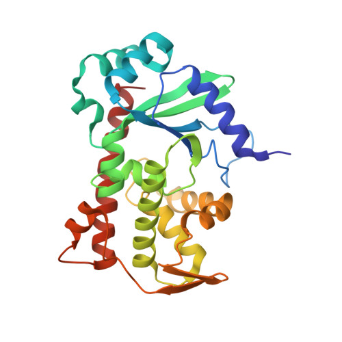
Crystal structure of Tam41 cytidine diphosphate diacylglycerol synthase from a Firmicutes bacterium.

Kimura, K.Kawai, F.Kubota-Kawai, H.Watanabe, Y.Tomii, K.Kojima, R.Hirata, K.Yamamori, Y.Endo, T.Tamura, Y.

(2022) J Biochem 171: 429-441

  • DOI: https://doi.org/10.1093/jb/mvab154
  • Primary Citation of Related Structures:  
    7ECD

  • PubMed Abstract: 

    Translocator assembly and maintenance 41 (Tam41) catalyses the synthesis of cytidine diphosphate diacylglycerol (CDP-DAG), which is a high-energy intermediate phospholipid critical for generating cardiolipin in mitochondria. Although Tam41 is present almost exclusively in eukaryotic cells, a Firmicutes bacterium contains the gene encoding Tam41-type CDP-DAG synthase (FbTam41). FbTam41 converted phosphatidic acid (PA) to CDP-DAG using a ternary complex mechanism in vitro. Additionally, FbTam41 functionally substituted yeast Tam41 in vivo. These results demonstrate that Tam41-type CDP-DAG synthase functions in some prokaryotic cells. We determined the crystal structure of FbTam41 lacking the C-terminal 18 residues in the cytidine triphosphate (CTP)-Mg2+ bound form at a resolution of 2.6 Å. The crystal structure showed that FbTam41 contained a positively charged pocket that specifically accommodated CTP-Mg2+ and PA in close proximity. By using this structure, we constructed a model for the full-length structure of FbTam41 containing the last a-helix, which was missing in the crystal structure. Based on this model, we propose a molecular mechanism for CDP-DAG synthesis in bacterial cells and mitochondria.


  • Organizational Affiliation

    Graduate School of Global Symbiotic Sciences, Yamagata University, 1-4-12 Kojirakawa-machi, Yamagata 990-8560, Japan.


Macromolecules
Find similar proteins by:  (by identity cutoff)  |  3D Structure
Entity ID: 1
MoleculeChains Sequence LengthOrganismDetailsImage
Phosphatidate cytidylyltransferase272Firmicutes bacterium CAG:884Mutation(s): 0 
Gene Names: BN804_00706
EC: 2.7.7.41
UniProt
Find proteins for R5MX27 (Firmicutes bacterium CAG:884)
Explore R5MX27 
Go to UniProtKB:  R5MX27
Entity Groups  
Sequence Clusters30% Identity50% Identity70% Identity90% Identity95% Identity100% Identity
UniProt GroupR5MX27
Sequence Annotations
Expand
  • Reference Sequence
Experimental Data & Validation

Experimental Data

  • Method: X-RAY DIFFRACTION
  • Resolution: 2.60 Å
  • R-Value Free: 0.238 
  • R-Value Work: 0.201 
  • R-Value Observed: 0.203 
  • Space Group: I 41
Unit Cell:
Length ( Å )Angle ( ˚ )
a = 104.159α = 90
b = 104.159β = 90
c = 113.959γ = 90
Software Package:
Software NamePurpose
PHENIXrefinement
XDSdata reduction
XDSdata scaling
PHENIXphasing

Structure Validation

View Full Validation Report


Ligand Structure Quality Assessment 


Entry History & Funding Information

Deposition Data


Funding OrganizationLocationGrant Number
Japan Society for the Promotion of Science (JSPS)Japan17H06414
Japan Society for the Promotion of Science (JSPS)Japan22227003
Japan Agency for Medical Research and Development (AMED)JapanJP20gm5910026
Japan Agency for Medical Research and Development (AMED)JapanJP20am0101070

Revision History  (Full details and data files)

  • Version 1.0: 2022-01-19
    Type: Initial release
  • Version 1.1: 2022-04-13
    Changes: Database references