7E5Q

Crystal Structure of Dye Decolorizing peroxidase from Bacillus subtilis at acidic pH


Experimental Data Snapshot

  • Method: X-RAY DIFFRACTION
  • Resolution: 1.90 Å
  • R-Value Free: 0.233 
  • R-Value Work: 0.180 
  • R-Value Observed: 0.182 

wwPDB Validation   3D Report Full Report


Ligand Structure Quality Assessment 


This is version 1.2 of the entry. See complete history


Literature

Structural insights at acidic pH of dye-decolorizing peroxidase from Bacillus subtilis.

Dhankhar, P.Dalal, V.Sharma, A.K.Kumar, P.

(2022) Proteins 

  • DOI: https://doi.org/10.1002/prot.26444
  • Primary Citation of Related Structures:  
    7E5Q

  • PubMed Abstract: 

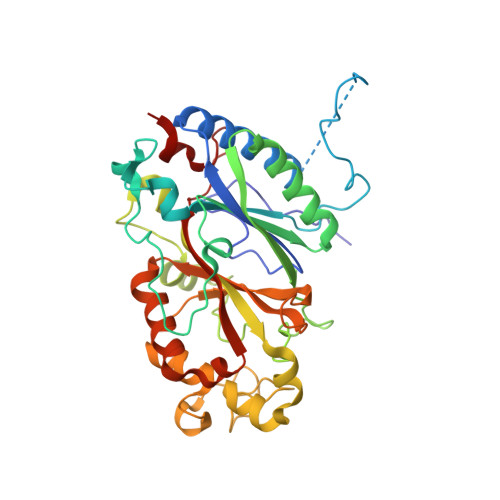
    Dye-decolorizing peroxidases (DyPs), a type of heme-containing oxidoreductase enzymes, catalyze the peroxide-dependent oxidation of various industrial dyes as well as lignin and lignin model compounds. In our previous work, we have recently reported the crystal structures of class A-type DyP from Bacillus subtilis at pH 7.0 (BsDyP7), exposing the location of three binding sites for small substrates and high redox-potential substrates. The biochemical studies revealed the optimum acidic pH for enzyme activity. In the present study, the crystal structure of BsDyP at acidic pH (BsDyP4) reveals two-monomer units stabilized by intermolecular salt bridges and a hydrogen bond network in a homo-dimeric unit. Based on the monomeric structural comparison of BsDyP4 and BsDyP7, minor differences were observed in the loop regions, that is, LI (Ala64-Gln71), LII (Glu96-Lys108), LIII (Pro117-Leu124), and LIV (Leu295-Asp303). Despite these differences, BsDyP4 adopts similar heme architecture as well as three substrate-binding sites to BsDyP7. In BsDyP4, a shift in Asp187, heme pocket residue discloses the plausible reason for optimal acidic pH for BsDyP activity. This study provides insight into the structural changes in BsDyP at acidic pH, where BsDyP is biologically active.


  • Organizational Affiliation

    Department of Biosciences and Bioengineering, Indian Institute of Technology Roorkee, Roorkee, India.


Macromolecules
Find similar proteins by:  (by identity cutoff)  |  3D Structure
Entity ID: 1
MoleculeChains Sequence LengthOrganismDetailsImage
Deferrochelatase/peroxidase
A, B
363Bacillus subtilisMutation(s): 0 
Gene Names: efeBGS595_16230
EC: 1.11.1
UniProt
Find proteins for P39597 (Bacillus subtilis (strain 168))
Explore P39597 
Go to UniProtKB:  P39597
Entity Groups  
Sequence Clusters30% Identity50% Identity70% Identity90% Identity95% Identity100% Identity
UniProt GroupP39597
Sequence Annotations
Expand
  • Reference Sequence
Small Molecules
Ligands 8 Unique
IDChains Name / Formula / InChI Key2D Diagram3D Interactions
HEM
Query on HEM

Download Ideal Coordinates CCD File 
C [auth A],
Z [auth B]
PROTOPORPHYRIN IX CONTAINING FE
C34 H32 Fe N4 O4
KABFMIBPWCXCRK-RGGAHWMASA-L
CIT
Query on CIT

Download Ideal Coordinates CCD File 
JA [auth B],
N [auth A]
CITRIC ACID
C6 H8 O7
KRKNYBCHXYNGOX-UHFFFAOYSA-N
PEG
Query on PEG

Download Ideal Coordinates CCD File 
O [auth A],
P [auth A],
Q [auth A]
DI(HYDROXYETHYL)ETHER
C4 H10 O3
MTHSVFCYNBDYFN-UHFFFAOYSA-N
GOL
Query on GOL

Download Ideal Coordinates CCD File 
KA [auth B]GLYCEROL
C3 H8 O3
PEDCQBHIVMGVHV-UHFFFAOYSA-N
EDO
Query on EDO

Download Ideal Coordinates CCD File 
BA [auth B]
CA [auth B]
DA [auth B]
E [auth A]
EA [auth B]
BA [auth B],
CA [auth B],
DA [auth B],
E [auth A],
EA [auth B],
F [auth A],
FA [auth B],
G [auth A],
GA [auth B],
H [auth A],
HA [auth B],
I [auth A],
IA [auth B],
J [auth A],
K [auth A],
L [auth A],
M [auth A],
Y [auth B]
1,2-ETHANEDIOL
C2 H6 O2
LYCAIKOWRPUZTN-UHFFFAOYSA-N
CL
Query on CL

Download Ideal Coordinates CCD File 
S [auth A]
SA [auth B]
T [auth A]
TA [auth B]
U [auth A]
S [auth A],
SA [auth B],
T [auth A],
TA [auth B],
U [auth A],
UA [auth B],
V [auth A],
W [auth A],
X [auth A]
CHLORIDE ION
Cl
VEXZGXHMUGYJMC-UHFFFAOYSA-M
OXY
Query on OXY

Download Ideal Coordinates CCD File 
AA [auth B],
D [auth A]
OXYGEN MOLECULE
O2
MYMOFIZGZYHOMD-UHFFFAOYSA-N
NA
Query on NA

Download Ideal Coordinates CCD File 
LA [auth B]
MA [auth B]
NA [auth B]
OA [auth B]
PA [auth B]
LA [auth B],
MA [auth B],
NA [auth B],
OA [auth B],
PA [auth B],
QA [auth B],
R [auth A],
RA [auth B]
SODIUM ION
Na
FKNQFGJONOIPTF-UHFFFAOYSA-N
Experimental Data & Validation

Experimental Data

  • Method: X-RAY DIFFRACTION
  • Resolution: 1.90 Å
  • R-Value Free: 0.233 
  • R-Value Work: 0.180 
  • R-Value Observed: 0.182 
  • Space Group: P 21 21 21
Unit Cell:
Length ( Å )Angle ( ˚ )
a = 40.855α = 90
b = 117.342β = 90
c = 135.441γ = 90
Software Package:
Software NamePurpose
PHENIXrefinement
PDB_EXTRACTdata extraction
Cootmodel building
PHASERphasing

Structure Validation

View Full Validation Report


Ligand Structure Quality Assessment 


Entry History & Funding Information

Deposition Data


Funding OrganizationLocationGrant Number
Department of Biotechnology (DBT, India)IndiaDBT-1088-BIO

Revision History  (Full details and data files)

  • Version 1.0: 2022-08-24
    Type: Initial release
  • Version 1.1: 2022-11-30
    Changes: Database references
  • Version 1.2: 2023-11-29
    Changes: Data collection, Refinement description