7CMJ

Crystal structure of L.donovani Hypoxanthine-guanine phosphoribosyl transferase (HGPRT)


Experimental Data Snapshot

  • Method: X-RAY DIFFRACTION
  • Resolution: 2.76 Å
  • R-Value Free: 0.225 
  • R-Value Work: 0.181 
  • R-Value Observed: 0.183 

wwPDB Validation   3D Report Full Report


Ligand Structure Quality Assessment 


This is version 1.2 of the entry. See complete history


Literature

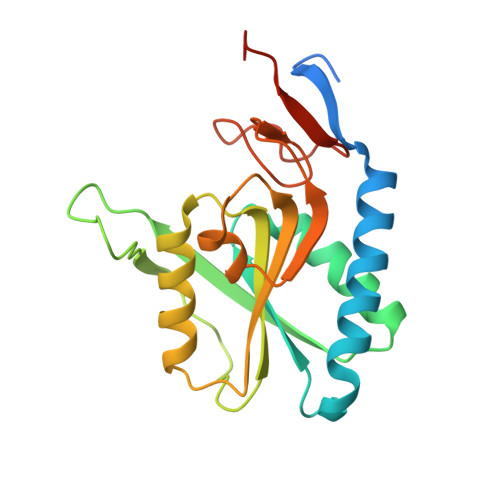
The L.donovani Hypoxanthine-guanine phosphoribosyl transferase (HGPRT) oligomer is distinct from the human homolog.

Parihar, P.S.Pratap, J.V.

(2020) Biochem Biophys Res Commun 532: 499-504

  • DOI: https://doi.org/10.1016/j.bbrc.2020.08.052
  • Primary Citation of Related Structures:  
    7CMJ

  • PubMed Abstract: 

    Purine bases, synthesized de novo or recycled through the salvage pathway, are precursors of nucleotide synthesis and are essential in a variety of physiological processes including cell division, growth, signaling, energy metabolism and synthesis of vitamins/co-factor. The protozoan kinetoplastid parasites including Leishmania cannot synthesize de novo and rely solely on the purine salvage pathway, recycling the degraded products of nucleic acid metabolism. Enzymes of this pathway are thus of therapeutic importance. The enzyme Hypoxanthine-guanine phosphoribosyl transferase (HGPRT) (EC 2.4.2.8) plays a central role in this pathway, converting the purine base to its monophosphate product. Towards the elucidation of its role, we have cloned, expressed, purified and determined the crystal structure of L. donovani HGPRT at 2.76 Å. Comparative structural analysis with the human homolog indicates differences in oligomer association. Comparative analyses identify insertions in the human homolog sequence in the tetramer interface. The results suggest that this difference can be exploited for therapeutic approaches.


  • Organizational Affiliation

    Molecular and Structural Biology Division, CSIR-Central Drug Research Institute, B.S. 10/1, Sector 10, Jankipuram Extension, Sitapur Road, Lucknow, 226031, U.P, India.


Macromolecules
Find similar proteins by:  (by identity cutoff)  |  3D Structure
Entity ID: 1
MoleculeChains Sequence LengthOrganismDetailsImage
Hypoxanthine phosphoribosyltransferase
A, B
211Leishmania donovani BPK282A1Mutation(s): 0 
Gene Names: LDBPK_210980
EC: 2.4.2.8
UniProt
Find proteins for Q7KLF3 (Leishmania donovani)
Explore Q7KLF3 
Go to UniProtKB:  Q7KLF3
Entity Groups  
Sequence Clusters30% Identity50% Identity70% Identity90% Identity95% Identity100% Identity
UniProt GroupQ7KLF3
Sequence Annotations
Expand
  • Reference Sequence
Small Molecules
Ligands 6 Unique
IDChains Name / Formula / InChI Key2D Diagram3D Interactions
PG4 (Subject of Investigation/LOI)
Query on PG4

Download Ideal Coordinates CCD File 
K [auth B]TETRAETHYLENE GLYCOL
C8 H18 O5
UWHCKJMYHZGTIT-UHFFFAOYSA-N
BA (Subject of Investigation/LOI)
Query on BA

Download Ideal Coordinates CCD File 
D [auth A],
I [auth B]
BARIUM ION
Ba
XDFCIPNJCBUZJN-UHFFFAOYSA-N
PEG (Subject of Investigation/LOI)
Query on PEG

Download Ideal Coordinates CCD File 
F [auth A],
G [auth B]
DI(HYDROXYETHYL)ETHER
C4 H10 O3
MTHSVFCYNBDYFN-UHFFFAOYSA-N
PO4 (Subject of Investigation/LOI)
Query on PO4

Download Ideal Coordinates CCD File 
E [auth A],
J [auth B]
PHOSPHATE ION
O4 P
NBIIXXVUZAFLBC-UHFFFAOYSA-K
CL (Subject of Investigation/LOI)
Query on CL

Download Ideal Coordinates CCD File 
L [auth B],
M [auth B]
CHLORIDE ION
Cl
VEXZGXHMUGYJMC-UHFFFAOYSA-M
MG (Subject of Investigation/LOI)
Query on MG

Download Ideal Coordinates CCD File 
C [auth A],
H [auth B]
MAGNESIUM ION
Mg
JLVVSXFLKOJNIY-UHFFFAOYSA-N
Experimental Data & Validation

Experimental Data

  • Method: X-RAY DIFFRACTION
  • Resolution: 2.76 Å
  • R-Value Free: 0.225 
  • R-Value Work: 0.181 
  • R-Value Observed: 0.183 
  • Space Group: P 65 2 2
Unit Cell:
Length ( Å )Angle ( ˚ )
a = 80.8α = 90
b = 80.8β = 90
c = 344.321γ = 120
Software Package:
Software NamePurpose
PHENIXrefinement
iMOSFLMdata reduction
Aimlessdata scaling
PHASERphasing

Structure Validation

View Full Validation Report


Ligand Structure Quality Assessment 


Entry History 

Deposition Data

Revision History  (Full details and data files)

  • Version 1.0: 2021-08-04
    Type: Initial release
  • Version 1.1: 2023-02-15
    Changes: Database references
  • Version 1.2: 2023-11-29
    Changes: Data collection, Refinement description