7BY0

The cryo-EM structure of CENP-A nucleosome in complex with the phosphorylated CENP-C


Experimental Data Snapshot

  • Method: ELECTRON MICROSCOPY
  • Resolution: 4.50 Å
  • Aggregation State: PARTICLE 
  • Reconstruction Method: SINGLE PARTICLE 

wwPDB Validation   3D Report Full Report


This is version 1.1 of the entry. See complete history


Literature

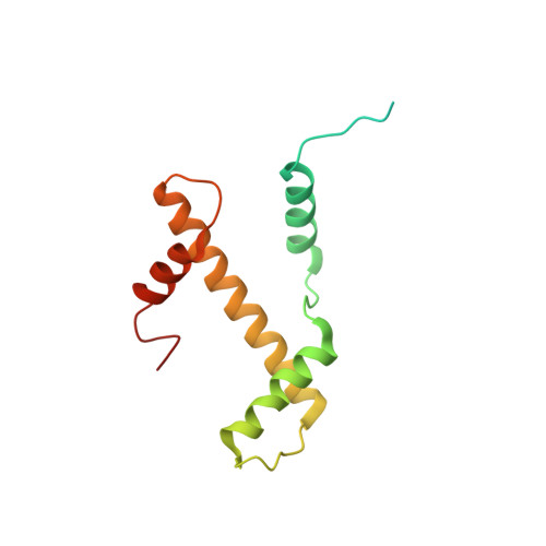
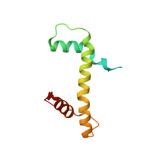
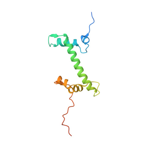
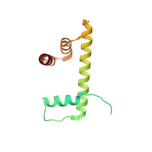
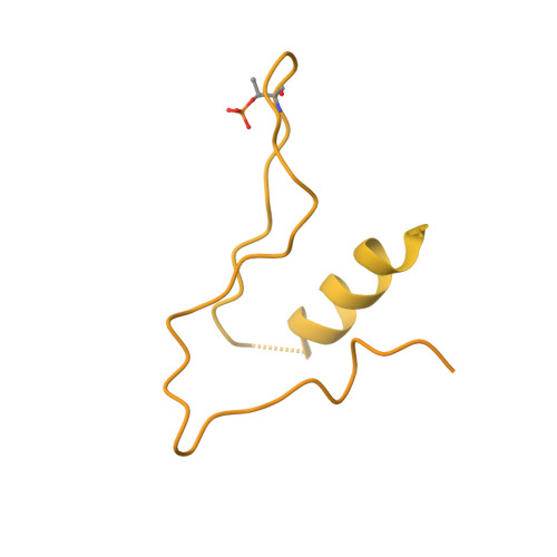
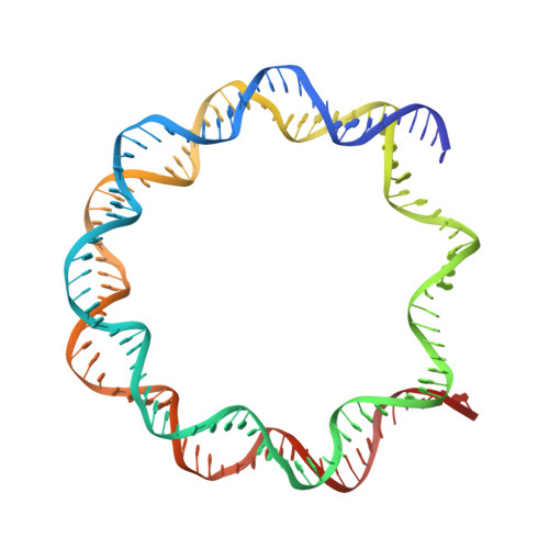
Cryo-EM structure of the CENP-A nucleosome in complex with phosphorylated CENP-C.

Ariyoshi, M.Makino, F.Watanabe, R.Nakagawa, R.Kato, T.Namba, K.Arimura, Y.Fujita, R.Kurumizaka, H.Okumura, E.I.Hara, M.Fukagawa, T.

(2021) EMBO J 40: e105671-e105671

  • DOI: https://doi.org/10.15252/embj.2020105671
  • Primary Citation of Related Structures:  
    7BXT, 7BY0

  • PubMed Abstract: 

    The CENP-A nucleosome is a key structure for kinetochore assembly. Once the CENP-A nucleosome is established in the centromere, additional proteins recognize the CENP-A nucleosome to form a kinetochore. CENP-C and CENP-N are CENP-A binding proteins. We previously demonstrated that vertebrate CENP-C binding to the CENP-A nucleosome is regulated by CDK1-mediated CENP-C phosphorylation. However, it is still unknown how the phosphorylation of CENP-C regulates its binding to CENP-A. It is also not completely understood how and whether CENP-C and CENP-N act together on the CENP-A nucleosome. Here, using cryo-electron microscopy (cryo-EM) in combination with biochemical approaches, we reveal a stable CENP-A nucleosome-binding mode of CENP-C through unique regions. The chicken CENP-C structure bound to the CENP-A nucleosome is stabilized by an intramolecular link through the phosphorylated CENP-C residue. The stable CENP-A-CENP-C complex excludes CENP-N from the CENP-A nucleosome. These findings provide mechanistic insights into the dynamic kinetochore assembly regulated by CDK1-mediated CENP-C phosphorylation.


  • Organizational Affiliation

    Graduate School of Frontier Biosciences, Osaka University, Suita, Osaka, Japan.


Macromolecules

Find similar proteins by:  (by identity cutoff)  |  3D Structure
Entity ID: 1
MoleculeChains Sequence LengthOrganismDetailsImage
Histone H3.2,Histone H3-like centromeric protein A
A, E
141Gallus gallusMutation(s): 0 
UniProt
Find proteins for Q6XXM1 (Gallus gallus)
Explore Q6XXM1 
Go to UniProtKB:  Q6XXM1
Find proteins for P84229 (Gallus gallus)
Explore P84229 
Go to UniProtKB:  P84229
Entity Groups  
Sequence Clusters30% Identity50% Identity70% Identity90% Identity95% Identity100% Identity
UniProt GroupsP84229Q6XXM1
Sequence Annotations
Expand
  • Reference Sequence
Find similar proteins by:  (by identity cutoff)  |  3D Structure
Entity ID: 2
MoleculeChains Sequence LengthOrganismDetailsImage
Histone H4
B, F
102Homo sapiensMutation(s): 0 
UniProt & NIH Common Fund Data Resources
Find proteins for P62805 (Homo sapiens)
Explore P62805 
Go to UniProtKB:  P62805
PHAROS:  P62805
Entity Groups  
Sequence Clusters30% Identity50% Identity70% Identity90% Identity95% Identity100% Identity
UniProt GroupP62805
Sequence Annotations
Expand
  • Reference Sequence
Find similar proteins by:  (by identity cutoff)  |  3D Structure
Entity ID: 3
MoleculeChains Sequence LengthOrganismDetailsImage
Histone H2A type 1-B/E
C, G
130Homo sapiensMutation(s): 0 
UniProt & NIH Common Fund Data Resources
Find proteins for P04908 (Homo sapiens)
Explore P04908 
Go to UniProtKB:  P04908
PHAROS:  P04908
Entity Groups  
Sequence Clusters30% Identity50% Identity70% Identity90% Identity95% Identity100% Identity
UniProt GroupP04908
Sequence Annotations
Expand
  • Reference Sequence
Find similar proteins by:  (by identity cutoff)  |  3D Structure
Entity ID: 4
MoleculeChains Sequence LengthOrganismDetailsImage
Histone H2B type 1-J
D, H
126Homo sapiensMutation(s): 0 
Gene Names: HIST1H2BJH2BFR
UniProt & NIH Common Fund Data Resources
Find proteins for P06899 (Homo sapiens)
Explore P06899 
Go to UniProtKB:  P06899
PHAROS:  P06899
GTEx:  ENSG00000124635 
Entity Groups  
Sequence Clusters30% Identity50% Identity70% Identity90% Identity95% Identity100% Identity
UniProt GroupP06899
Sequence Annotations
Expand
  • Reference Sequence
Find similar proteins by:  (by identity cutoff)  |  3D Structure
Entity ID: 7
MoleculeChains Sequence LengthOrganismDetailsImage
Maltose Binding Protein tag, linker,CENP-C
K, L
664unidentified cloning vectorGallus gallus
This entity is chimeric
Mutation(s): 0 
UniProt
Find proteins for P0AEX9 (Escherichia coli (strain K12))
Explore P0AEX9 
Go to UniProtKB:  P0AEX9
Find proteins for O57393 (Gallus gallus)
Explore O57393 
Go to UniProtKB:  O57393
Entity Groups  
Sequence Clusters30% Identity50% Identity70% Identity90% Identity95% Identity100% Identity
UniProt GroupsO57393P0AEX9
Sequence Annotations
Expand
  • Reference Sequence
Find similar nucleic acids by:  (by identity cutoff)  |  3D Structure
Entity ID: 5
MoleculeChains LengthOrganismImage
DNA (145-MER)145synthetic construct
Sequence Annotations
Expand
  • Reference Sequence
Find similar nucleic acids by:  (by identity cutoff)  |  3D Structure
Entity ID: 6
MoleculeChains LengthOrganismImage
DNA (145-MER)145synthetic construct
Sequence Annotations
Expand
  • Reference Sequence
Small Molecules
Modified Residues  1 Unique
IDChains TypeFormula2D DiagramParent
TPO
Query on TPO
K, L
L-PEPTIDE LINKINGC4 H10 N O6 PTHR
Experimental Data & Validation

Experimental Data

  • Method: ELECTRON MICROSCOPY
  • Resolution: 4.50 Å
  • Aggregation State: PARTICLE 
  • Reconstruction Method: SINGLE PARTICLE 
EM Software:
TaskSoftware PackageVersion
RECONSTRUCTIONRELION3.08

Structure Validation

View Full Validation Report


Entry History & Funding Information

Deposition Data


Funding OrganizationLocationGrant Number
Japan Society for the Promotion of Science (JSPS)Japan25221106
Japan Agency for Medical Research and Development (AMED)Japan0101117
Japan Agency for Medical Research and Development (AMED)Japan0101020

Revision History  (Full details and data files)

  • Version 1.0: 2021-02-10
    Type: Initial release
  • Version 1.1: 2021-03-10
    Changes: Database references