How Coiled-Coil Assemblies Accommodate Multiple Aromatic Residues.
Rhys, G.G., Dawson, W.M., Beesley, J.L., Martin, F.J.O., Brady, R.L., Thomson, A.R., Woolfson, D.N.(2021) Biomacromolecules 22: 2010-2019
- PubMed: 33881308 Search on PubMed
- DOI: https://doi.org/10.1021/acs.biomac.1c00131
- Primary Citation Related Structures:
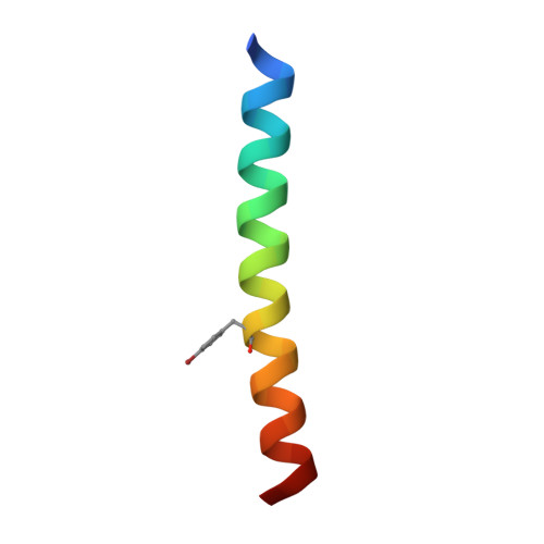
7BO8, 7BO9, 7BOA - PubMed Abstract:
Rational protein design requires understanding the contribution of each amino acid to a targeted protein fold. For a subset of protein structures, namely, α-helical coiled coils (CCs), knowledge is sufficiently advanced to allow the rational de novo design of many structures, including entirely new protein folds. Current CC design rules center on using aliphatic hydrophobic residues predominantly to drive the folding and assembly of amphipathic α helices. The consequences of using aromatic residues-which would be useful for introducing structural probes, and binding and catalytic functionalities-into these interfaces are not understood. There are specific examples of designed CCs containing such aromatic residues, e.g ., phenylalanine-rich sequences, and the use of polar aromatic residues to make buried hydrogen-bond networks. However, it is not known generally if sequences rich in tyrosine can form CCs, or what CC assemblies these would lead to. Here, we explore tyrosine-rich sequences in a general CC-forming background and resolve new CC structures. In one of these, an antiparallel tetramer, the tyrosine residues are solvent accessible and pack at the interface between the core and the surface. In another more complex structure, the residues are buried and form an extended hydrogen-bond network.
- School of Chemistry, University of Bristol, Cantock's Close, Bristol BS8 1TS, United Kingdom.
Organizational Affiliation: