7BAV

A de novo pentameric coiled-coil assembly: CC-Type2-(TgLaId)4-W19BrPhe.


Experimental Data Snapshot

  • Method: X-RAY DIFFRACTION
  • Resolution: 1.30 Å
  • R-Value Free: 0.214 
  • R-Value Work: 0.186 
  • R-Value Observed: 0.187 

wwPDB Validation   3D Report Full Report


This is version 1.1 of the entry. See complete history


Literature

Coiled coils 9-to-5: rational de novo design of alpha-helical barrels with tunable oligomeric states.

Dawson, W.M.Martin, F.J.O.Rhys, G.G.Shelley, K.L.Brady, R.L.Woolfson, D.N.

(2021) Chem Sci 12: 6923-6928

  • DOI: https://doi.org/10.1039/d1sc00460c
  • Primary Citation of Related Structures:  
    7A1T, 7BAS, 7BAT, 7BAU, 7BAV, 7BAW, 7BIM

  • PubMed Abstract: 

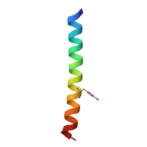
    The rational design of linear peptides that assemble controllably and predictably in water is challenging. Short sequences must encode unique target structures and avoid alternative states. However, the non-covalent forces that stabilize and discriminate between states are weak. Nonetheless, for α-helical coiled-coil assemblies considerable progress has been made in rational de novo design. In these, sequence repeats of nominally hydrophobic ( h ) and polar ( p ) residues, hpphppp , direct the assembly of amphipathic helices into dimeric to tetrameric bundles. Expanding this pattern to hpphhph can produce larger α-helical barrels. Here, we show that pentameric to nonameric barrels are accessed by varying the residue at one of the h sites. In peptides with four L/I-K-E-I-A-x-Z repeats, decreasing the size of Z from threonine to serine to alanine to glycine gives progressively larger oligomers. X-ray crystal structures of the resulting α-helical barrels rationalize this: side chains at Z point directly into the helical interfaces, and smaller residues allow closer helix contacts and larger assemblies.


  • Organizational Affiliation

    School of Chemistry, University of Bristol Cantock's Close Bristol BS8 1TS UK w.dawson@bristol.ac.uk d.n.woolfson@bristol.ac.uk.


Macromolecules
Find similar proteins by:  (by identity cutoff)  |  3D Structure
Entity ID: 1
MoleculeChains Sequence LengthOrganismDetailsImage
CC-Type2-(TgLaId)4-W19BrPhe
A, B, C, D, E
32synthetic constructMutation(s): 0 
Entity Groups  
Sequence Clusters30% Identity50% Identity70% Identity90% Identity95% Identity100% Identity
Sequence Annotations
Expand
  • Reference Sequence
Small Molecules
Modified Residues  1 Unique
IDChains TypeFormula2D DiagramParent
4BF
Query on 4BF
A, B, C, D, E
L-PEPTIDE LINKINGC9 H10 Br N O2TYR
Experimental Data & Validation

Experimental Data

  • Method: X-RAY DIFFRACTION
  • Resolution: 1.30 Å
  • R-Value Free: 0.214 
  • R-Value Work: 0.186 
  • R-Value Observed: 0.187 
  • Space Group: P 21 21 21
Unit Cell:
Length ( Å )Angle ( ˚ )
a = 43.734α = 90
b = 51.831β = 90
c = 55.04γ = 90
Software Package:
Software NamePurpose
Aimlessdata scaling
REFMACrefinement
PDB_EXTRACTdata extraction
DIALSdata reduction
ACORNphasing

Structure Validation

View Full Validation Report


Entry History & Funding Information

Deposition Data


Funding OrganizationLocationGrant Number
European Research Council (ERC)United Kingdom340764
Engineering and Physical Sciences Research CouncilUnited KingdomEP/G036764
Biotechnology and Biological Sciences Research Council (BBSRC)United KingdomBB R00661X 1

Revision History  (Full details and data files)

  • Version 1.0: 2021-04-28
    Type: Initial release
  • Version 1.1: 2021-11-17
    Changes: Data collection, Database references, Structure summary