7B7Z

DeAMPylation complex of monomeric FICD and AMPylated BiP (state 1)


Experimental Data Snapshot

  • Method: X-RAY DIFFRACTION
  • Resolution: 1.70 Å
  • R-Value Free: 0.221 
  • R-Value Work: 0.195 
  • R-Value Observed: 0.196 

wwPDB Validation   3D Report Full Report


This is version 1.2 of the entry. See complete history


Literature

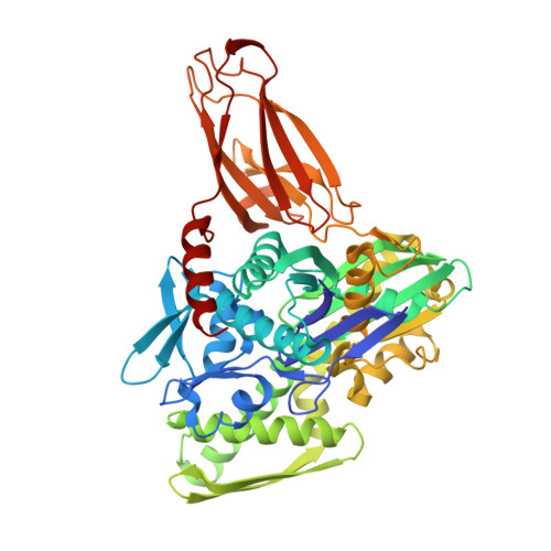
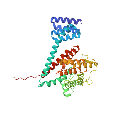
Structures of a deAMPylation complex rationalise the switch between antagonistic catalytic activities of FICD.

Perera, L.A.Preissler, S.Zaccai, N.R.Prevost, S.Devos, J.M.Haertlein, M.Ron, D.

(2021) Nat Commun 12: 5004-5004

  • DOI: https://doi.org/10.1038/s41467-021-25076-7
  • Primary Citation of Related Structures:  
    7B7Z, 7B80

  • PubMed Abstract: 

    The endoplasmic reticulum (ER) Hsp70 chaperone BiP is regulated by AMPylation, a reversible inactivating post-translational modification. Both BiP AMPylation and deAMPylation are catalysed by a single ER-localised enzyme, FICD. Here we present crystallographic and solution structures of a deAMPylation Michaelis complex formed between mammalian AMPylated BiP and FICD. The latter, via its tetratricopeptide repeat domain, binds a surface that is specific to ATP-state Hsp70 chaperones, explaining the exquisite selectivity of FICD for BiP's ATP-bound conformation both when AMPylating and deAMPylating Thr518. The eukaryotic deAMPylation mechanism thus revealed, rationalises the role of the conserved Fic domain Glu234 as a gatekeeper residue that both inhibits AMPylation and facilitates hydrolytic deAMPylation catalysed by dimeric FICD. These findings point to a monomerisation-induced increase in Glu234 flexibility as the basis of an oligomeric state-dependent switch between FICD's antagonistic activities, despite a similar mode of engagement of its two substrates - unmodified and AMPylated BiP.


  • Organizational Affiliation

    Cambridge Institute for Medical Research, University of Cambridge, Cambridge, UK. lp397@cam.ac.uk.


Macromolecules
Find similar proteins by:  (by identity cutoff)  |  3D Structure
Entity ID: 1
MoleculeChains Sequence LengthOrganismDetailsImage
Endoplasmic reticulum chaperone BiPA [auth B]523Cricetulus griseusMutation(s): 2 
Gene Names: HSPA5GRP78I79_019946
EC: 3.6.4.10
UniProt
Find proteins for G3I8R9 (Cricetulus griseus)
Explore G3I8R9 
Go to UniProtKB:  G3I8R9
Entity Groups  
Sequence Clusters30% Identity50% Identity70% Identity90% Identity95% Identity100% Identity
UniProt GroupG3I8R9
Sequence Annotations
Expand
  • Reference Sequence
Find similar proteins by:  (by identity cutoff)  |  3D Structure
Entity ID: 2
MoleculeChains Sequence LengthOrganismDetailsImage
Protein adenylyltransferase FICDB [auth A]343Homo sapiensMutation(s): 2 
Gene Names: FICDHIP13HYPEUNQ3041/PRO9857
EC: 2.7.7 (PDB Primary Data), 3.1.4 (PDB Primary Data)
UniProt & NIH Common Fund Data Resources
Find proteins for Q9BVA6 (Homo sapiens)
Explore Q9BVA6 
Go to UniProtKB:  Q9BVA6
PHAROS:  Q9BVA6
GTEx:  ENSG00000198855 
Entity Groups  
Sequence Clusters30% Identity50% Identity70% Identity90% Identity95% Identity100% Identity
UniProt GroupQ9BVA6
Sequence Annotations
Expand
  • Reference Sequence
Experimental Data & Validation

Experimental Data

  • Method: X-RAY DIFFRACTION
  • Resolution: 1.70 Å
  • R-Value Free: 0.221 
  • R-Value Work: 0.195 
  • R-Value Observed: 0.196 
  • Space Group: P 21 21 21
Unit Cell:
Length ( Å )Angle ( ˚ )
a = 95.371α = 90
b = 104.076β = 90
c = 105.629γ = 90
Software Package:
Software NamePurpose
REFMACrefinement
PDB_EXTRACTdata extraction
DIALSdata reduction
Aimlessdata scaling
PHASERphasing

Structure Validation

View Full Validation Report


Entry History & Funding Information

Deposition Data


Funding OrganizationLocationGrant Number
Wellcome TrustUnited Kingdom200848/Z/16/Z

Revision History  (Full details and data files)

  • Version 1.0: 2021-07-07
    Type: Initial release
  • Version 1.1: 2021-10-06
    Changes: Data collection, Database references
  • Version 1.2: 2024-01-31
    Changes: Data collection, Refinement description