6XPC

Structure of human GGT1 in complex with full GSH molecule


Experimental Data Snapshot

  • Method: X-RAY DIFFRACTION
  • Resolution: 2.26 Å
  • R-Value Free: 0.210 
  • R-Value Work: 0.159 
  • R-Value Observed: 0.162 

wwPDB Validation   3D Report Full Report


Ligand Structure Quality Assessment 


This is version 1.2 of the entry. See complete history


Literature

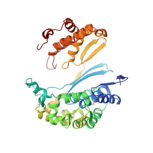
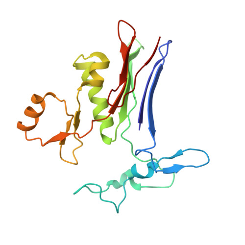
Crystal structures of glutathione- and inhibitor-bound human GGT1: critical interactions within the cysteinylglycine binding site.

Terzyan, S.S.Nguyen, L.T.Burgett, A.W.G.Heroux, A.Smith, C.A.You, Y.Hanigan, M.H.

(2020) J Biol Chem 296: 100066-100066

  • DOI: https://doi.org/10.1074/jbc.RA120.016265
  • Primary Citation of Related Structures:  
    6XPB, 6XPC

  • PubMed Abstract: 

    Overexpression of γ-glutamyl transpeptidase (GGT1) has been implicated in an array of human diseases including asthma, reperfusion injury, and cancer. Inhibitors are needed for therapy, but development of potent, specific inhibitors of GGT1 has been hampered by a lack of structural information regarding substrate binding and cleavage. To enhance our understanding of the molecular mechanism of substrate cleavage, we have solved the crystal structures of human GGT1 (hGGT1) with glutathione (a substrate) and a phosphate-glutathione analog (an irreversible inhibitor) bound in the active site. These are the first structures of any eukaryotic GGT with the cysteinylglycine region of the substrate-binding site occupied. These structures and the structure of apo-hGGT reveal movement of amino acid residues within the active site as the substrate binds. Asn-401 and Thr-381 each form hydrogen bonds with two atoms of GSH spanning the γ-glutamyl bond. Three different atoms of hGGT1 interact with the carboxyl oxygen of the cysteine of GSH. Interactions between the enzyme and substrate change as the substrate moves deeper into the active site cleft. The substrate reorients and a new hydrogen bond is formed between the substrate and the oxyanion hole. Thr-381 is locked into a single conformation as an acyl bond forms between the substrate and the enzyme. These data provide insight on a molecular level into the substrate specificity of hGGT1 and provide an explanation for seemingly disparate observations regarding the enzymatic activity of hGGT1 mutants. This knowledge will aid in the design of clinically useful hGGT1 inhibitors.


  • Organizational Affiliation

    Laboratory of Biomolecular Structure and Function, Department of Biochemistry and Molecular Biology, University of Oklahoma Health Sciences Center, Oklahoma City, Oklahoma, USA.


Macromolecules
Find similar proteins by:  (by identity cutoff)  |  3D Structure
Entity ID: 1
MoleculeChains Sequence LengthOrganismDetailsImage
Glutathione hydrolase 1 heavy chain353Homo sapiensMutation(s): 0 
Gene Names: GGT1GGT
EC: 3.4.19.13 (PDB Primary Data), 2.3.2.2 (PDB Primary Data), 3.4.19.14 (PDB Primary Data)
UniProt & NIH Common Fund Data Resources
Find proteins for P19440 (Homo sapiens)
Explore P19440 
Go to UniProtKB:  P19440
PHAROS:  P19440
GTEx:  ENSG00000100031 
Entity Groups  
Sequence Clusters30% Identity50% Identity70% Identity90% Identity95% Identity100% Identity
UniProt GroupP19440
Sequence Annotations
Expand
  • Reference Sequence
Find similar proteins by:  (by identity cutoff)  |  3D Structure
Entity ID: 2
MoleculeChains Sequence LengthOrganismDetailsImage
Glutathione hydrolase 1 light chain189Homo sapiensMutation(s): 0 
Gene Names: GGT1GGT
EC: 3.4.19.13 (PDB Primary Data), 2.3.2.2 (PDB Primary Data), 3.4.19.14 (PDB Primary Data)
UniProt & NIH Common Fund Data Resources
Find proteins for P19440 (Homo sapiens)
Explore P19440 
Go to UniProtKB:  P19440
PHAROS:  P19440
GTEx:  ENSG00000100031 
Entity Groups  
Sequence Clusters30% Identity50% Identity70% Identity90% Identity95% Identity100% Identity
UniProt GroupP19440
Sequence Annotations
Expand
  • Reference Sequence
Small Molecules
Ligands 4 Unique
IDChains Name / Formula / InChI Key2D Diagram3D Interactions
GSH (Subject of Investigation/LOI)
Query on GSH

Download Ideal Coordinates CCD File 
L [auth B]GLUTATHIONE
C10 H17 N3 O6 S
RWSXRVCMGQZWBV-WDSKDSINSA-N
NAG
Query on NAG

Download Ideal Coordinates CCD File 
C [auth A]
D [auth A]
E [auth A]
F [auth A]
G [auth A]
C [auth A],
D [auth A],
E [auth A],
F [auth A],
G [auth A],
J [auth B]
2-acetamido-2-deoxy-beta-D-glucopyranose
C8 H15 N O6
OVRNDRQMDRJTHS-FMDGEEDCSA-N
CL
Query on CL

Download Ideal Coordinates CCD File 
H [auth A],
K [auth B]
CHLORIDE ION
Cl
VEXZGXHMUGYJMC-UHFFFAOYSA-M
NA
Query on NA

Download Ideal Coordinates CCD File 
I [auth A]SODIUM ION
Na
FKNQFGJONOIPTF-UHFFFAOYSA-N
Experimental Data & Validation

Experimental Data

  • Method: X-RAY DIFFRACTION
  • Resolution: 2.26 Å
  • R-Value Free: 0.210 
  • R-Value Work: 0.159 
  • R-Value Observed: 0.162 
  • Space Group: C 2 2 21
Unit Cell:
Length ( Å )Angle ( ˚ )
a = 105.395α = 90
b = 124.679β = 90
c = 104.989γ = 90
Software Package:
Software NamePurpose
REFMACrefinement
XDSdata reduction
Aimlessdata scaling
PDB_EXTRACTdata extraction
REFMACphasing

Structure Validation

View Full Validation Report


Ligand Structure Quality Assessment 


Entry History & Funding Information

Deposition Data


Funding OrganizationLocationGrant Number
National Institutes of Health/National Institute of General Medical Sciences (NIH/NIGMS)United StatesR01GM125952

Revision History  (Full details and data files)

  • Version 1.0: 2020-11-25
    Type: Initial release
  • Version 1.1: 2021-07-21
    Changes: Database references
  • Version 1.2: 2023-10-18
    Changes: Data collection, Database references, Refinement description