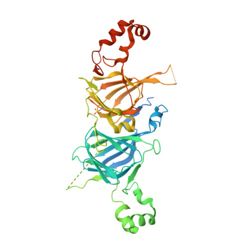
6V7G

Binding of Benzoic Acid and Anions Within the Cupin Domains of the Vicillin Protein Canavalin from Jack Bean (canavalia ensiformis): Crystal Structures


Experimental Data Snapshot

  • Method: X-RAY DIFFRACTION
  • Resolution: 1.40 Å
  • R-Value Free: 0.188 
  • R-Value Work: 0.160 
  • R-Value Observed: 0.161 

wwPDB Validation   3D Report Full Report


Ligand Structure Quality Assessment 


This is version 1.3 of the entry. See complete history


Literature

Binding of benzoic acid and anions within the cupin domains of the vicilin protein canavalin from jack bean (Canavalia ensiformis): Crystal structures.

McPherson, A.

(2020) Biochem Biophys Res Commun 524: 268-271

  • DOI: https://doi.org/10.1016/j.bbrc.2020.01.101
  • Primary Citation of Related Structures:  
    6V7G, 6V7J, 6V7L

  • PubMed Abstract: 

    X-ray intensities extending to 1.4 Å resolution were collected on the P6 3 hexagonal crystal form of canavalin, and extended to 1.9 Å for the orthorhombic C222 1 crystals. Structure determination of a new crystal form of canavalin having space group P2 1 2 1 2 1 is reported as well. Both the N and C terminal cupin domains contained identifiable ligands. For hexagonal crystals, in the cavity of the C terminal cupin, a molecule of benzoic acid was found, bound through carboxyl oxygens to Histidine 297, asparagine 284 and Arginine 376. The benzene ring was immersed in a cluster of at least 8 hydrophobic amino acid side chains. The N terminal cupin contained a molecule of citrate. Benzoic acid was also found to be present in the C terminal cupins of in the C222 1 and P2 1 2 1 2 1 crystal forms. In rhombohedral crystals, the C terminal cupin domain appeared to be occupied by a phosphate ion, but this was ambiguous. In cubic crystals, both domains were vacant. The N terminal cupin domains of canavalin in the P2 1 2 1 2 1 and rhombohedral crystals were also vacant, but the N terminal cupin domain of the C222 1 crystals contained a ligand whose identity is uncertain, but which has been modeled as HEPES buffer. A possible physiological role for the ligands and their complexes with canavalin is considered.


  • Organizational Affiliation

    Dept. Molecular Biology and Biochemistry, University of California, Irvine, 3205 McGaugh Hall, Irvine, CA, 92697, USA. Electronic address: amcphers@uci.edu.


Macromolecules
Find similar proteins by:  (by identity cutoff)  |  3D Structure
Entity ID: 1
MoleculeChains Sequence LengthOrganismDetailsImage
Canavalin445Canavalia ensiformisMutation(s): 0 
UniProt
Find proteins for P50477 (Canavalia ensiformis)
Explore P50477 
Go to UniProtKB:  P50477
Entity Groups  
Sequence Clusters30% Identity50% Identity70% Identity90% Identity95% Identity100% Identity
UniProt GroupP50477
Sequence Annotations
Expand
  • Reference Sequence
Small Molecules
Modified Residues  1 Unique
IDChains TypeFormula2D DiagramParent
OCS
Query on OCS
A
L-PEPTIDE LINKINGC3 H7 N O5 SCYS
Experimental Data & Validation

Experimental Data

  • Method: X-RAY DIFFRACTION
  • Resolution: 1.40 Å
  • R-Value Free: 0.188 
  • R-Value Work: 0.160 
  • R-Value Observed: 0.161 
  • Space Group: P 63
Unit Cell:
Length ( Å )Angle ( ˚ )
a = 125.806α = 90
b = 125.806β = 90
c = 49.875γ = 120
Software Package:
Software NamePurpose
REFMACrefinement
XDSdata reduction
Aimlessdata scaling
PHASERphasing

Structure Validation

View Full Validation Report


Ligand Structure Quality Assessment 


Entry History 

Deposition Data

  • Released Date: 2020-02-19 
  • Deposition Author(s): McPherson, A.
  • This entry supersedes: 6CB4

Revision History  (Full details and data files)

  • Version 1.0: 2020-02-19
    Type: Initial release
  • Version 1.1: 2020-03-18
    Changes: Database references
  • Version 1.2: 2023-10-11
    Changes: Data collection, Database references, Refinement description
  • Version 1.3: 2023-11-15
    Changes: Data collection