Binding of benzoic acid and anions within the cupin domains of the vicilin protein canavalin from jack bean (Canavalia ensiformis): Crystal structures.
McPherson, A.(2020) Biochem Biophys Res Commun 524: 268-271
- PubMed: 31983433 Search on PubMed
- DOI: https://doi.org/10.1016/j.bbrc.2020.01.101
- Primary Citation Related Structures:
6V7G, 6V7J, 6V7L - PubMed Abstract:
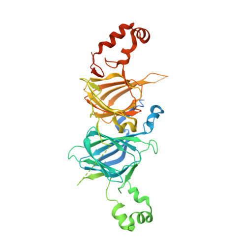
X-ray intensities extending to 1.4 Å resolution were collected on the P6 3 hexagonal crystal form of canavalin, and extended to 1.9 Å for the orthorhombic C222 1 crystals. Structure determination of a new crystal form of canavalin having space group P2 1 2 1 2 1 is reported as well. Both the N and C terminal cupin domains contained identifiable ligands. For hexagonal crystals, in the cavity of the C terminal cupin, a molecule of benzoic acid was found, bound through carboxyl oxygens to Histidine 297, asparagine 284 and Arginine 376. The benzene ring was immersed in a cluster of at least 8 hydrophobic amino acid side chains. The N terminal cupin contained a molecule of citrate. Benzoic acid was also found to be present in the C terminal cupins of in the C222 1 and P2 1 2 1 2 1 crystal forms. In rhombohedral crystals, the C terminal cupin domain appeared to be occupied by a phosphate ion, but this was ambiguous. In cubic crystals, both domains were vacant. The N terminal cupin domains of canavalin in the P2 1 2 1 2 1 and rhombohedral crystals were also vacant, but the N terminal cupin domain of the C222 1 crystals contained a ligand whose identity is uncertain, but which has been modeled as HEPES buffer. A possible physiological role for the ligands and their complexes with canavalin is considered.
- Dept. Molecular Biology and Biochemistry, University of California, Irvine, 3205 McGaugh Hall, Irvine, CA, 92697, USA. Electronic address: amcphers@uci.edu.
Organizational Affiliation: