6T13

CRYSTAL STRUCTURE OF GLUCOCEREBROSIDASE IN COMPLEX WITH A PYRROLOPYRAZINE


Experimental Data Snapshot

  • Method: X-RAY DIFFRACTION
  • Resolution: 1.85 Å
  • R-Value Free: 0.204 
  • R-Value Work: 0.174 
  • R-Value Observed: 0.176 

wwPDB Validation   3D Report Full Report


Ligand Structure Quality Assessment 


This is version 1.2 of the entry. See complete history


Literature

Novel beta-Glucocerebrosidase Activators That Bind to a New Pocket at a Dimer Interface and Induce Dimerization.

Benz, J.Rufer, A.C.Huber, S.Ehler, A.Hug, M.Topp, A.Guba, W.Hofmann, E.C.Jagasia, R.Rodriguez Sarmiento, R.M.

(2021) Angew Chem Int Ed Engl 60: 5436-5442

  • DOI: https://doi.org/10.1002/anie.202013890
  • Primary Citation of Related Structures:  
    6T13

  • PubMed Abstract: 

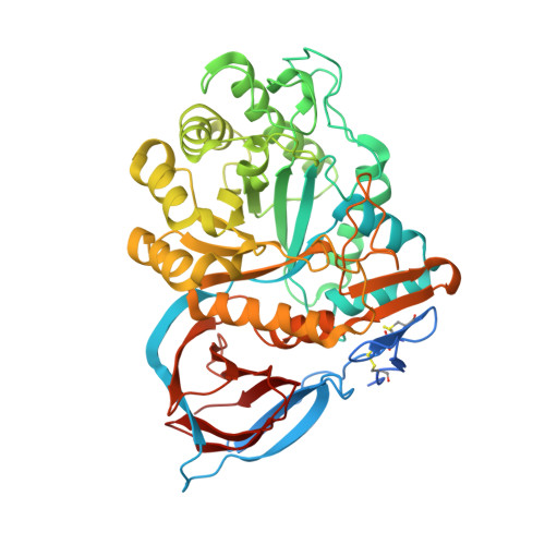
    Genetic, preclinical and clinical data link Parkinson's disease and Gaucher's disease and provide a rational entry point to disease modification therapy via enhancement of β-Glucocerebrosidase (GCase) activity. We discovered a new class of pyrrolo[2,3-b]pyrazine activators effecting both Vmax and Km. They bind to human GCase and increase substrate metabolism in the lysosome in a cellular assay. We obtained the first crystal structure for an activator and identified a novel non-inhibitory binding mode at the interface of a dimer, rationalizing the observed structure-activity relationship (SAR). The compound binds GCase inducing formation of a dimeric state at both endoplasmic reticulum (ER) and lysosomal pHs, as confirmed by analytical ultracentrifugation. Importantly, the pyrrolo[2,3-b]pyrazines have central nervous system (CNS) drug-like properties. Our findings are important for future drug discovery efforts in the field of GCase activation and provide a deeper mechanistic understanding of the requirements for enzymatic activation, pointing to the relevance of dimerization.


  • Organizational Affiliation

    Lead Discovery, Roche Innovation Center Basel, F. Hoffmann-La Roche Ltd., Grenzacherstrasse 124, 4070, Basel, Switzerland.


Macromolecules
Find similar proteins by:  (by identity cutoff)  |  3D Structure
Entity ID: 1
MoleculeChains Sequence LengthOrganismDetailsImage
Glucosylceramidase
A, B, C, D
536Homo sapiensMutation(s): 0 
EC: 3.2.1.45
UniProt & NIH Common Fund Data Resources
Find proteins for P04062 (Homo sapiens)
Explore P04062 
Go to UniProtKB:  P04062
PHAROS:  P04062
GTEx:  ENSG00000177628 
Entity Groups  
Sequence Clusters30% Identity50% Identity70% Identity90% Identity95% Identity100% Identity
UniProt GroupP04062
Sequence Annotations
Expand
  • Reference Sequence
Oligosaccharides

Help

Entity ID: 2
MoleculeChains Length2D Diagram Glycosylation3D Interactions
2-acetamido-2-deoxy-beta-D-glucopyranose-(1-4)-2-acetamido-2-deoxy-beta-D-glucopyranose
E
2N-Glycosylation
Glycosylation Resources
GlyTouCan:  G42666HT
GlyCosmos:  G42666HT
GlyGen:  G42666HT
Small Molecules
Ligands 7 Unique
IDChains Name / Formula / InChI Key2D Diagram3D Interactions
M7H (Subject of Investigation/LOI)
Query on M7H

Download Ideal Coordinates CCD File 
AA [auth C],
FA [auth D]
1-[4-[2-(4-methoxyphenyl)-5-methyl-pyrrolo[2,3-b]pyrazin-6-yl]piperidin-1-yl]ethanone
C21 H24 N4 O2
HEAGFUMRBLFGAD-UHFFFAOYSA-N
NAG
Query on NAG

Download Ideal Coordinates CCD File 
EA [auth D],
N [auth B],
Z [auth C]
2-acetamido-2-deoxy-beta-D-glucopyranose
C8 H15 N O6
OVRNDRQMDRJTHS-FMDGEEDCSA-N
TRS
Query on TRS

Download Ideal Coordinates CCD File 
J [auth A],
T [auth B]
2-AMINO-2-HYDROXYMETHYL-PROPANE-1,3-DIOL
C4 H12 N O3
LENZDBCJOHFCAS-UHFFFAOYSA-O
SO4
Query on SO4

Download Ideal Coordinates CCD File 
BA [auth C]
CA [auth C]
F [auth A]
G [auth A]
GA [auth D]
BA [auth C],
CA [auth C],
F [auth A],
G [auth A],
GA [auth D],
H [auth A],
HA [auth D],
I [auth A],
IA [auth D],
JA [auth D],
P [auth B],
Q [auth B],
R [auth B],
S [auth B]
SULFATE ION
O4 S
QAOWNCQODCNURD-UHFFFAOYSA-L
GOL
Query on GOL

Download Ideal Coordinates CCD File 
O [auth B]GLYCEROL
C3 H8 O3
PEDCQBHIVMGVHV-UHFFFAOYSA-N
EDO
Query on EDO

Download Ideal Coordinates CCD File 
K [auth A]
L [auth A]
LA [auth D]
M [auth A]
U [auth B]
K [auth A],
L [auth A],
LA [auth D],
M [auth A],
U [auth B],
V [auth B],
W [auth B],
X [auth B],
Y [auth B]
1,2-ETHANEDIOL
C2 H6 O2
LYCAIKOWRPUZTN-UHFFFAOYSA-N
CL
Query on CL

Download Ideal Coordinates CCD File 
DA [auth C],
KA [auth D]
CHLORIDE ION
Cl
VEXZGXHMUGYJMC-UHFFFAOYSA-M
Experimental Data & Validation

Experimental Data

  • Method: X-RAY DIFFRACTION
  • Resolution: 1.85 Å
  • R-Value Free: 0.204 
  • R-Value Work: 0.174 
  • R-Value Observed: 0.176 
  • Space Group: P 21 21 21
Unit Cell:
Length ( Å )Angle ( ˚ )
a = 90.849α = 90
b = 135.408β = 90
c = 191.384γ = 90
Software Package:
Software NamePurpose
XDSdata reduction
XPREPdata scaling
PHASERphasing
BUSTERrefinement
PDB_EXTRACTdata extraction

Structure Validation

View Full Validation Report


Ligand Structure Quality Assessment 


Entry History 

Deposition Data

Revision History  (Full details and data files)

  • Version 1.0: 2020-12-23
    Type: Initial release
  • Version 1.1: 2021-07-21
    Changes: Database references
  • Version 1.2: 2024-01-24
    Changes: Data collection, Database references, Refinement description