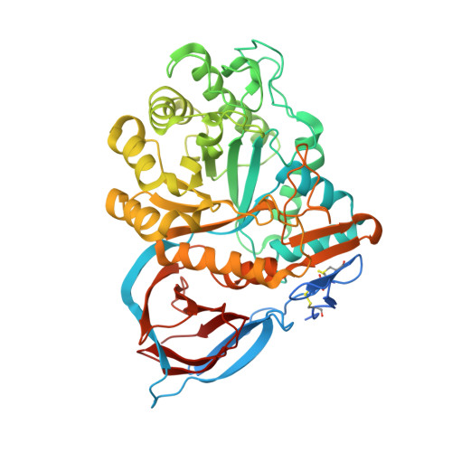
6T13

CRYSTAL STRUCTURE OF GLUCOCEREBROSIDASE IN COMPLEX WITH A PYRROLOPYRAZINE


Experimental Data Snapshot

  • Method: X-RAY DIFFRACTION
  • Resolution: 1.85 Å
  • R-Value Free: 0.204 
  • R-Value Work: 0.174 
  • R-Value Observed: 0.176 

wwPDB Validation   3D Report Full Report


Ligand Structure Quality Assessment 


This is version 1.1 of the entry. See complete history


Literature

Novel beta-Glucocerebrosidase Activators That Bind to a New Pocket at a Dimer Interface and Induce Dimerization.

Benz, J.Rufer, A.C.Huber, S.Ehler, A.Hug, M.Topp, A.Guba, W.Hofmann, E.C.Jagasia, R.Rodriguez Sarmiento, R.M.

(2021) Angew Chem Int Ed Engl 60: 5436-5442

  • DOI: https://doi.org/10.1002/anie.202013890
  • Primary Citation of Related Structures:  
    6T13

  • PubMed Abstract: 
  • Genetic, preclinical and clinical data link Parkinson's disease and Gaucher's disease and provide a rational entry point to disease modification therapy via enhancement of β-Glucocerebrosidase (GCase) activity. We discovered a new class of pyrrolo[2,3-b]pyrazine activators effecting both Vmax and Km ...

    Genetic, preclinical and clinical data link Parkinson's disease and Gaucher's disease and provide a rational entry point to disease modification therapy via enhancement of β-Glucocerebrosidase (GCase) activity. We discovered a new class of pyrrolo[2,3-b]pyrazine activators effecting both Vmax and Km. They bind to human GCase and increase substrate metabolism in the lysosome in a cellular assay. We obtained the first crystal structure for an activator and identified a novel non-inhibitory binding mode at the interface of a dimer, rationalizing the observed structure-activity relationship (SAR). The compound binds GCase inducing formation of a dimeric state at both endoplasmic reticulum (ER) and lysosomal pHs, as confirmed by analytical ultracentrifugation. Importantly, the pyrrolo[2,3-b]pyrazines have central nervous system (CNS) drug-like properties. Our findings are important for future drug discovery efforts in the field of GCase activation and provide a deeper mechanistic understanding of the requirements for enzymatic activation, pointing to the relevance of dimerization.


    Organizational Affiliation

    Medicinal Chemistry, Roche Pharma Research and Early Development (pRED), Roche Innovation Center Basel, F. Hoffmann-La Roche Ltd., Grenzacherstrasse 124, 4070, Basel, Switzerland.


Macromolecules
Find similar proteins by:  (by identity cutoff)  |  3D Structure
Entity ID: 1
MoleculeChainsSequence LengthOrganismDetailsImage
GlucosylceramidaseA, B, C, D536Homo sapiensMutation(s): 0 
Gene Names: GBAGCGLUC
EC: 3.2.1.45 (PDB Primary Data), 2.4.1 (UniProt), 3.2.1 (UniProt), 3.2.1.46 (UniProt)
UniProt & NIH Common Fund Data Resources
Find proteins for P04062 (Homo sapiens)
Explore P04062 
Go to UniProtKB:  P04062
PHAROS:  P04062
Entity Groups  
Sequence Clusters30% Identity50% Identity70% Identity90% Identity95% Identity100% Identity
UniProt GroupP04062
Protein Feature View
Expand
  • Reference Sequence
Oligosaccharides

Help

Entity ID: 2
MoleculeChainsChain Length2D DiagramGlycosylation3D Interactions
2-acetamido-2-deoxy-beta-D-glucopyranose-(1-4)-2-acetamido-2-deoxy-beta-D-glucopyranoseE 2N-Glycosylation Oligosaccharides Interaction
Glycosylation Resources
GlyTouCan:  G42666HT
GlyCosmos:  G42666HT
GlyGen:  G42666HT
Small Molecules
Ligands 7 Unique
IDChainsName / Formula / InChI Key2D Diagram3D Interactions
M7H (Subject of Investigation/LOI)
Query on M7H

Download Ideal Coordinates CCD File 
AA [auth C],
FA [auth D]
1-[4-[2-(4-methoxyphenyl)-5-methyl-pyrrolo[2,3-b]pyrazin-6-yl]piperidin-1-yl]ethanone
C21 H24 N4 O2
HEAGFUMRBLFGAD-UHFFFAOYSA-N
 Ligand Interaction
NAG
Query on NAG

Download Ideal Coordinates CCD File 
EA [auth D],
N [auth B],
Z [auth C]
2-acetamido-2-deoxy-beta-D-glucopyranose
C8 H15 N O6
OVRNDRQMDRJTHS-FMDGEEDCSA-N
 Ligand Interaction
TRS
Query on TRS

Download Ideal Coordinates CCD File 
J [auth A],
T [auth B]
2-AMINO-2-HYDROXYMETHYL-PROPANE-1,3-DIOL
C4 H12 N O3
LENZDBCJOHFCAS-UHFFFAOYSA-O
 Ligand Interaction
SO4
Query on SO4

Download Ideal Coordinates CCD File 
BA [auth C],
CA [auth C],
F [auth A],
G [auth A],
GA [auth D],
BA [auth C],
CA [auth C],
F [auth A],
G [auth A],
GA [auth D],
H [auth A],
HA [auth D],
I [auth A],
IA [auth D],
JA [auth D],
P [auth B],
Q [auth B],
R [auth B],
S [auth B]
SULFATE ION
O4 S
QAOWNCQODCNURD-UHFFFAOYSA-L
 Ligand Interaction
GOL
Query on GOL

Download Ideal Coordinates CCD File 
O [auth B]GLYCEROL
C3 H8 O3
PEDCQBHIVMGVHV-UHFFFAOYSA-N
 Ligand Interaction
EDO
Query on EDO

Download Ideal Coordinates CCD File 
K [auth A],
L [auth A],
LA [auth D],
M [auth A],
U [auth B],
K [auth A],
L [auth A],
LA [auth D],
M [auth A],
U [auth B],
V [auth B],
W [auth B],
X [auth B],
Y [auth B]
1,2-ETHANEDIOL
C2 H6 O2
LYCAIKOWRPUZTN-UHFFFAOYSA-N
 Ligand Interaction
CL
Query on CL

Download Ideal Coordinates CCD File 
DA [auth C],
KA [auth D]
CHLORIDE ION
Cl
VEXZGXHMUGYJMC-UHFFFAOYSA-M
 Ligand Interaction
Experimental Data & Validation

Experimental Data

  • Method: X-RAY DIFFRACTION
  • Resolution: 1.85 Å
  • R-Value Free: 0.204 
  • R-Value Work: 0.174 
  • R-Value Observed: 0.176 
  • Space Group: P 21 21 21
Unit Cell:
Length ( Å )Angle ( ˚ )
a = 90.849α = 90
b = 135.408β = 90
c = 191.384γ = 90
Software Package:
Software NamePurpose
XDSdata reduction
XPREPdata scaling
PHASERphasing
BUSTERrefinement
PDB_EXTRACTdata extraction

Structure Validation

View Full Validation Report


Ligand Structure Quality Assessment 


Entry History 

Deposition Data

Revision History  (Full details and data files)

  • Version 1.0: 2020-12-23
    Type: Initial release
  • Version 1.1: 2021-07-21
    Changes: Database references