6GAU

Extremely 'open' clamp structure of DNA gyrase: role of the Corynebacteriales GyrB specific insert


Experimental Data Snapshot

  • Method: X-RAY DIFFRACTION
  • Resolution: 3.30 Å
  • R-Value Free: 0.251 
  • R-Value Work: 0.173 
  • R-Value Observed: 0.177 

wwPDB Validation   3D Report Full Report


Ligand Structure Quality Assessment 


This is version 1.3 of the entry. See complete history


Literature

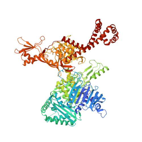
Overall Structures of Mycobacterium tuberculosis DNA Gyrase Reveal the Role of a Corynebacteriales GyrB-Specific Insert in ATPase Activity.

Petrella, S.Capton, E.Raynal, B.Giffard, C.Thureau, A.Bonnete, F.Alzari, P.M.Aubry, A.Mayer, C.

(2019) Structure 27: 579-589.e5

  • DOI: https://doi.org/10.1016/j.str.2019.01.004
  • Primary Citation of Related Structures:  
    6GAU, 6GAV

  • PubMed Abstract: 
  • Despite sharing common features, previous studies have shown that gyrases from different species have been modified throughout evolution to modulate their properties. Here, we report two crystal structures of Mycobacterium tuberculosis DNA gyrase, an apo and AMPPNP-bound form at 2 ...

    Despite sharing common features, previous studies have shown that gyrases from different species have been modified throughout evolution to modulate their properties. Here, we report two crystal structures of Mycobacterium tuberculosis DNA gyrase, an apo and AMPPNP-bound form at 2.6-Å and 3.3-Å resolution, respectively. These structures provide high-resolution structural data on the quaternary organization and interdomain connections of a gyrase (full-length GyrB-GyrA57) 2 thus providing crucial inputs on this essential drug target. Together with small-angle X-ray scattering studies, they revealed an "extremely open" N-gate state, which persists even in the DNA-free gyrase-AMPPNP complex and an unexpected connection between the ATPase and cleavage core domains mediated by two Corynebacteriales-specific motifs, respectively the C-loop and DEEE-loop. We show that the C-loop participates in the stabilization of this open conformation, explaining why this gyrase has a lower ATPase activity. Our results image a conformational state which might be targeted for drug discovery.


    Organizational Affiliation

    Unité de Microbiologie Structurale, Institut Pasteur, CNRS UMR 3528, 25 rue du Docteur Roux, 75724 Paris Cedex 15, France; Université Paris Diderot, Sorbonne Paris Cité, 75724 Paris Cedex 15, France.


Macromolecules
Find similar proteins by:  (by identity cutoff)  |  3D Structure
Entity ID: 1
MoleculeChainsSequence LengthOrganismDetailsImage
DNA gyrase subunit B,DNA gyrase subunit A
A, B
1,179Mycobacterium tuberculosisMutation(s): 0 
Gene Names: gyrBgyrARv0006MTCY10H4.04
EC: 5.99.1.3 (PDB Primary Data), 5.6.2.2 (UniProt)
UniProt
Find proteins for F6N7X0 (Mycobacterium tuberculosis)
Explore F6N7X0 
Go to UniProtKB:  F6N7X0
Find proteins for P9WG47 (Mycobacterium tuberculosis (strain ATCC 25618 / H37Rv))
Explore P9WG47 
Go to UniProtKB:  P9WG47
Entity Groups  
Sequence Clusters30% Identity50% Identity70% Identity90% Identity95% Identity100% Identity
UniProt GroupsF6N7X0P9WG47
Protein Feature View
Expand
  • Reference Sequence
Small Molecules
Ligands 2 Unique
IDChainsName / Formula / InChI Key2D Diagram3D Interactions
ANP
Query on ANP

Download Ideal Coordinates CCD File 
C [auth A],
E [auth B]
PHOSPHOAMINOPHOSPHONIC ACID-ADENYLATE ESTER
C10 H17 N6 O12 P3
PVKSNHVPLWYQGJ-KQYNXXCUSA-N
 Ligand Interaction
MG
Query on MG

Download Ideal Coordinates CCD File 
D [auth A],
F [auth B]
MAGNESIUM ION
Mg
JLVVSXFLKOJNIY-UHFFFAOYSA-N
 Ligand Interaction
Experimental Data & Validation

Experimental Data

  • Method: X-RAY DIFFRACTION
  • Resolution: 3.30 Å
  • R-Value Free: 0.251 
  • R-Value Work: 0.173 
  • R-Value Observed: 0.177 
  • Space Group: P 1
Unit Cell:
Length ( Å )Angle ( ˚ )
a = 91.523α = 75.64
b = 96.782β = 64.44
c = 105.791γ = 65.8
Software Package:
Software NamePurpose
BUSTERrefinement
XSCALEdata scaling
PHASERphasing
PDB_EXTRACTdata extraction
XDSdata reduction

Structure Validation

View Full Validation Report


Ligand Structure Quality Assessment 


Entry History 

Deposition Data

Revision History  (Full details and data files)

  • Version 1.0: 2019-02-20
    Type: Initial release
  • Version 1.1: 2019-04-10
    Changes: Data collection, Database references
  • Version 1.2: 2019-05-08
    Changes: Advisory, Data collection, Derived calculations
  • Version 1.3: 2019-10-16
    Changes: Data collection