6CPL | pdb_00006cpl

Crystal structure of DR11 presenting the gag293 epitope


Experimental Data Snapshot

  • Method: X-RAY DIFFRACTION
  • Resolution: 2.45 Å
  • R-Value Free: 
    0.253 (Depositor), 0.257 (DCC) 
  • R-Value Work: 
    0.224 (Depositor), 0.224 (DCC) 
  • R-Value Observed: 
    0.225 (Depositor) 

Starting Model: experimental
View more details

wwPDB Validation 3D Report Full Report

Validation slider image for 6CPL

Ligand Structure Quality Assessment 


This is version 2.2 of the entry. See complete history

Literature

CD4+T cell-mediated HLA class II cross-restriction in HIV controllers.

Galperin, M.Farenc, C.Mukhopadhyay, M.Jayasinghe, D.Decroos, A.Benati, D.Tan, L.L.Ciacchi, L.Reid, H.H.Rossjohn, J.Chakrabarti, L.A.Gras, S.

(2018) Sci Immunol 3

  • DOI: https://doi.org/10.1126/sciimmunol.aat0687
  • Primary Citation Related Structures: 
    6CPH, 6CPL, 6CPN, 6CPO, 6CQJ, 6CQL, 6CQN, 6CQQ, 6CQR

  • PubMed Abstract: 

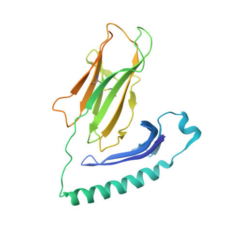
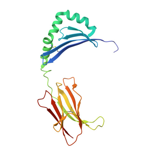
    Rare individuals, termed HIV controllers, spontaneously control HIV infection by mounting efficient T cell responses against the virus. Protective CD4 + T cell responses from HIV controllers involve high-affinity public T cell receptors (TCRs) recognizing an immunodominant capsid epitope (Gag293) presented by a remarkably broad array of human leukocyte antigen (HLA) class II molecules. Here, we determine the structures of a prototypical public TCR bound to HLA-DR1, HLA-DR11, and HLA-DR15 molecules presenting the Gag293 epitope. TCR recognition was driven by contacts with the Gag293 epitope, a feature that underpinned the extensive HLA cross-restriction. These high-affinity TCRs promoted mature immunological synapse formation and cytotoxic capacity in both CD4 + and CD8 + T cells. The public TCRs suppressed HIV replication in multiple genetic backgrounds ex vivo, emphasizing the functional advantage conferred by broad HLA class II cross-restriction.


  • Organizational Affiliation
    • Pasteur Institute, Viral Pathogenesis Unit, Paris, France.

Macromolecule Content 

  • Total Structure Weight: 52.32 kDa 
  • Atom Count: 3,462 
  • Modeled Residue Count: 385 
  • Deposited Residue Count: 439 
  • Unique protein chains: 3

Macromolecules

Find similar proteins by:|  3D Structure
Entity ID: 1
MoleculeChains  Sequence LengthOrganismDetailsImage
HLA class II histocompatibility antigen, DR alpha chain229Homo sapiensMutation(s): 0 
Gene Names: HLA-DRAHLA-DRA1
UniProt & NIH Common Fund Data Resources
Find proteins for P01903 (Homo sapiens)
Explore P01903 
Go to UniProtKB:  P01903
PHAROS:  P01903
GTEx:  ENSG00000204287 
Entity Groups
Sequence Clusters30% Identity50% Identity70% Identity90% Identity95% Identity100% Identity
UniProt GroupP01903
Glycosylation
Glycosylation Sites: 2Go to GlyGen: P01903-1
Sequence Annotations
Expand
Reference Sequence
Find similar proteins by:|  3D Structure
Entity ID: 2
MoleculeChains  Sequence LengthOrganismDetailsImage
HLA class II histocompatibility antigen, DRB1-11 beta chain190Homo sapiensMutation(s): 0 
Gene Names: HLA-DRB1
UniProt & NIH Common Fund Data Resources
Find proteins for P01911 (Homo sapiens)
Explore P01911 
Go to UniProtKB:  P01911
PHAROS:  P01911
GTEx:  ENSG00000196126 
Entity Groups
Sequence Clusters30% Identity50% Identity70% Identity90% Identity95% Identity100% Identity
UniProt GroupP01911
Glycosylation
Glycosylation Sites: 1Go to GlyGen: P01911-1
Sequence Annotations
Expand
Reference Sequence
Find similar proteins by:  Sequence   |   3D Structure  
Entity ID: 3
MoleculeChains  Sequence LengthOrganismDetailsImage
Gag polyprotein20HIV-1 M:B_HXB2RMutation(s): 0 
UniProt
Find proteins for P04585 (Human immunodeficiency virus type 1 group M subtype B (isolate HXB2))
Explore P04585 
Go to UniProtKB:  P04585
Entity Groups
Sequence Clusters30% Identity50% Identity70% Identity90% Identity95% Identity100% Identity
UniProt GroupP04585
Sequence Annotations
Expand
Reference Sequence

Oligosaccharides

Help  
Entity ID: 4
MoleculeChains Length2D Diagram GlycosylationD Interactions
2-acetamido-2-deoxy-beta-D-glucopyranose-(1-4)-2-acetamido-2-deoxy-beta-D-glucopyranose
D
2N-Glycosylation
Glycosylation Resources
GlyTouCan: G42666HT
GlyCosmos: G42666HT
GlyGen: G42666HT

Small Molecules

Ligands 4 Unique
IDChains Name / Formula / InChI Key2D Diagram3D Interactions
NAG

Query on NAG


Download:Ideal Coordinates CCD File
E [auth A],
I [auth B]
2-acetamido-2-deoxy-beta-D-glucopyranose
C8 H15 N O6
OVRNDRQMDRJTHS-FMDGEEDCSA-N
SO4

Query on SO4


Download:Ideal Coordinates CCD File
F [auth A],
G [auth A],
L [auth B],
M [auth B],
N [auth B]
SULFATE ION
O4 S
QAOWNCQODCNURD-UHFFFAOYSA-L
ACT

Query on ACT


Download:Ideal Coordinates CCD File
J [auth B],
K [auth B]
ACETATE ION
C2 H3 O2
QTBSBXVTEAMEQO-UHFFFAOYSA-M
NA

Query on NA


Download:Ideal Coordinates CCD File
H [auth A],
O [auth B],
P [auth C]
SODIUM ION
Na
FKNQFGJONOIPTF-UHFFFAOYSA-N

Experimental Data & Validation

Experimental Data

  • Method: X-RAY DIFFRACTION
  • Resolution: 2.45 Å
  • R-Value Free:  0.253 (Depositor), 0.257 (DCC) 
  • R-Value Work:  0.224 (Depositor), 0.224 (DCC) 
  • R-Value Observed: 0.225 (Depositor) 
Space Group: P 31 1 2
Unit Cell:
Length ( Å )Angle ( ˚ )
a = 65.746α = 90
b = 65.746β = 90
c = 222.514γ = 120
Software Package:
Software NamePurpose
BUSTERrefinement
XDSdata reduction
Aimlessdata scaling

Structure Validation

View Full Validation Report


Ligand Structure Quality Assessment 


Entry History 

Deposition Data

Revision History  (Full details and data files)

  • Version 1.0: 2018-06-06
    Type: Initial release
  • Version 1.1: 2018-06-27
    Changes: Data collection, Database references, Structure summary
  • Version 2.0: 2020-07-29
    Type: Remediation
    Reason: Carbohydrate remediation
    Changes: Atomic model, Data collection, Derived calculations, Structure summary
  • Version 2.1: 2023-10-04
    Changes: Data collection, Database references, Refinement description, Structure summary
  • Version 2.2: 2024-11-20
    Changes: Structure summary