6Z79

Variant Surface Glycoprotein VSGsur, I3C ("Magic Triangle") derivative used for phasing of the structure and subsequently as a model for molecular replacement of native, mutants, and drug soaks.


Experimental Data Snapshot

  • Method: X-RAY DIFFRACTION
  • Resolution: 1.92 Å
  • R-Value Free: 0.253 
  • R-Value Work: 0.234 
  • R-Value Observed: 0.235 

wwPDB Validation   3D Report Full Report


Ligand Structure Quality Assessment 


This is version 1.0 of the entry. See complete history


Literature

Structure of trypanosome coat protein VSGsur and function in suramin resistance.

Zeelen, J.van Straaten, M.Verdi, J.Hempelmann, A.Hashemi, H.Perez, K.Jeffrey, P.D.Halg, S.Wiedemar, N.Maser, P.Papavasiliou, F.N.Stebbins, C.E.

(2021) Nat Microbiol 6: 392-400

  • DOI: https://doi.org/10.1038/s41564-020-00844-1
  • Primary Citation of Related Structures:  
    6Z79, 6Z7B, 6Z7C, 6Z7D, 6Z7E, 6Z8G, 6Z8H

  • PubMed Abstract: 

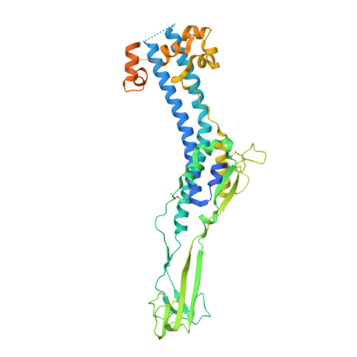
    Suramin has been a primary early-stage treatment for African trypanosomiasis for nearly 100 yr. Recent studies revealed that trypanosome strains that express the variant surface glycoprotein (VSG) VSGsur possess heightened resistance to suramin. Here, we show that VSGsur binds tightly to suramin but other VSGs do not. By solving high-resolution crystal structures of VSGsur and VSG13, we also demonstrate that these VSGs define a structurally divergent subgroup of the coat proteins. The co-crystal structure of VSGsur with suramin reveals that the chemically symmetric drug binds within a large cavity in the VSG homodimer asymmetrically, primarily through contacts of its central benzene rings. Structure-based, loss-of-contact mutations in VSGsur significantly decrease the affinity to suramin and lead to a loss of the resistance phenotype. Altogether, these data show that the resistance phenotype is dependent on the binding of suramin to VSGsur, establishing that the VSG proteins can possess functionality beyond their role in antigenic variation.


  • Organizational Affiliation

    Division of Structural Biology of Infection and Immunity, German Cancer Research Center, Heidelberg, Germany.


Macromolecules
Find similar proteins by:  (by identity cutoff)  |  3D Structure
Entity ID: 1
MoleculeChains Sequence LengthOrganismDetailsImage
Variant surface glycoprotein Sur491Trypanosoma brucei rhodesienseMutation(s): 0 
Gene Names: VSGsur
Membrane Entity: Yes 
UniProt
Find proteins for A0A291L8F4 (Trypanosoma brucei rhodesiense)
Explore A0A291L8F4 
Go to UniProtKB:  A0A291L8F4
Entity Groups  
Sequence Clusters30% Identity50% Identity70% Identity90% Identity95% Identity100% Identity
UniProt GroupA0A291L8F4
Sequence Annotations
Expand
  • Reference Sequence
Oligosaccharides

Help

Entity ID: 2
MoleculeChains Length2D Diagram Glycosylation3D Interactions
alpha-D-mannopyranose-(1-3)-[alpha-D-mannopyranose-(1-6)]beta-D-mannopyranose-(1-4)-2-acetamido-2-deoxy-beta-D-glucopyranose-(1-4)-2-acetamido-2-deoxy-beta-D-glucopyranose
B
5N-Glycosylation
Glycosylation Resources
GlyTouCan:  G22768VO
GlyCosmos:  G22768VO
GlyGen:  G22768VO
Small Molecules
Ligands 1 Unique
IDChains Name / Formula / InChI Key2D Diagram3D Interactions
I3C (Subject of Investigation/LOI)
Query on I3C

Download Ideal Coordinates CCD File 
C [auth A]5-amino-2,4,6-triiodobenzene-1,3-dicarboxylic acid
C8 H4 I3 N O4
JEZJSNULLBSYHV-UHFFFAOYSA-N
Experimental Data & Validation

Experimental Data

  • Method: X-RAY DIFFRACTION
  • Resolution: 1.92 Å
  • R-Value Free: 0.253 
  • R-Value Work: 0.234 
  • R-Value Observed: 0.235 
  • Space Group: P 21 21 2
Unit Cell:
Length ( Å )Angle ( ˚ )
a = 46.955α = 90
b = 70.893β = 90
c = 129.652γ = 90
Software Package:
Software NamePurpose
PHENIXrefinement
XDSdata processing
Cootmodel building
SHELXDEphasing

Structure Validation

View Full Validation Report


Ligand Structure Quality Assessment 


Entry History 

Deposition Data

Revision History  (Full details and data files)

  • Version 1.0: 2021-03-17
    Type: Initial release