6Y93

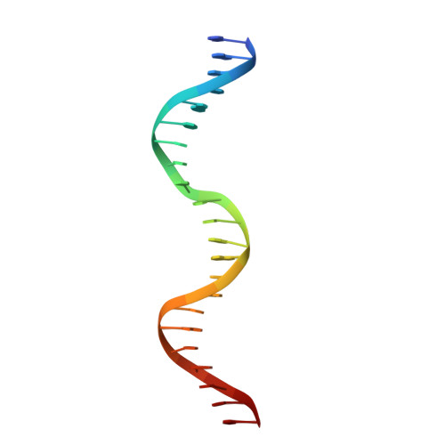
Crystal structure of the DNA-binding domain of the Nucleoid Occlusion Factor (Noc) complexed to the Noc-binding site (NBS)


Experimental Data Snapshot

  • Method: X-RAY DIFFRACTION
  • Resolution: 2.23 Å
  • R-Value Free: 0.279 
  • R-Value Work: 0.231 
  • R-Value Observed: 0.234 

wwPDB Validation   3D Report Full Report


This is version 1.0 of the entry. See complete history


Literature

Diversification of DNA-Binding Specificity by Permissive and Specificity-Switching Mutations in the ParB/Noc Protein Family.

Jalal, A.S.B.Tran, N.T.Stevenson, C.E.Chan, E.W.Lo, R.Tan, X.Noy, A.Lawson, D.M.Le, T.B.K.

(2020) Cell Rep 32: 107928-107928

  • DOI: 10.1016/j.celrep.2020.107928
  • Primary Citation of Related Structures:  
    6S6H, 6Y93

  • PubMed Abstract: 
  • Specific interactions between proteins and DNA are essential to many biological processes. Yet, it remains unclear how the diversification in DNA-binding specificity was brought about, and the mutational paths that led to changes in specificity are unknown ...

    Specific interactions between proteins and DNA are essential to many biological processes. Yet, it remains unclear how the diversification in DNA-binding specificity was brought about, and the mutational paths that led to changes in specificity are unknown. Using a pair of evolutionarily related DNA-binding proteins, each with a different DNA preference (ParB [Partitioning Protein B] and Noc [Nucleoid Occlusion Factor], which both play roles in bacterial chromosome maintenance), we show that specificity is encoded by a set of four residues at the protein-DNA interface. Combining X-ray crystallography and deep mutational scanning of the interface, we suggest that permissive mutations must be introduced before specificity-switching mutations to reprogram specificity and that mutational paths to new specificity do not necessarily involve dual-specificity intermediates. Overall, our results provide insight into the possible evolutionary history of ParB and Noc and, in a broader context, might be useful for understanding the evolution of other classes of DNA-binding proteins.


    Organizational Affiliation

    Department of Molecular Microbiology, John Innes Centre, Norwich NR4 7UH, UK. Electronic address: tung.le@jic.ac.uk.


Macromolecules

Find similar proteins by:  (by identity cutoff)  |  3D Structure
Entity ID: 1
MoleculeChainsSequence LengthOrganismDetailsImage
Nucleoid occlusion proteinA, B146Bacillus subtilis subsp. subtilis str. 168Mutation(s): 0 
Gene Names: nocyyaABSU40990
UniProt
Find proteins for P37524 (Bacillus subtilis (strain 168))
Explore P37524 
Go to UniProtKB:  P37524
Entity Groups  
Sequence Clusters30% Identity50% Identity70% Identity90% Identity95% Identity100% Identity
UniProt GroupP37524
Protein Feature View
Expand
  • Reference Sequence

Find similar nucleic acids by:  Sequence   |   3D Structure  

Entity ID: 2
MoleculeChainsLengthOrganismImage
Noc Binding Site (NBS)C, D22synthetic construct
Protein Feature View
Expand
  • Reference Sequence
Experimental Data & Validation

Experimental Data

  • Method: X-RAY DIFFRACTION
  • Resolution: 2.23 Å
  • R-Value Free: 0.279 
  • R-Value Work: 0.231 
  • R-Value Observed: 0.234 
  • Space Group: C 1 2 1
Unit Cell:
Length ( Å )Angle ( ˚ )
a = 134.139α = 90
b = 60.567β = 116.88
c = 81.053γ = 90
Software Package:
Software NamePurpose
STARANISOdata scaling
REFMACrefinement
PDB_EXTRACTdata extraction
DIALSdata reduction
PHASERphasing

Structure Validation

View Full Validation Report


Entry History & Funding Information

Deposition Data


Funding OrganizationLocationGrant Number
Royal SocietyUnited KingdomUF140053
Royal SocietyUnited KingdomRG150448
Biotechnology and Biological Sciences Research Council (BBSRC)United KingdomBB/P018165/1
Biotechnology and Biological Sciences Research Council (BBSRC)United KingdomBBS/E/J/000C0683
Biotechnology and Biological Sciences Research Council (BBSRC)United KingdomBBS/E/J/000PR9791

Revision History  (Full details and data files)

  • Version 1.0: 2020-08-05
    Type: Initial release