6S6H

Crystal structure of the DNA binding domain of the chromosome-partitioning protein ParB complexed to the centromeric parS site


Experimental Data Snapshot

  • Method: X-RAY DIFFRACTION
  • Resolution: 2.40 Å
  • R-Value Free: 0.232 
  • R-Value Work: 0.216 
  • R-Value Observed: 0.217 

wwPDB Validation   3D Report Full Report


This is version 1.2 of the entry. See complete history


Literature

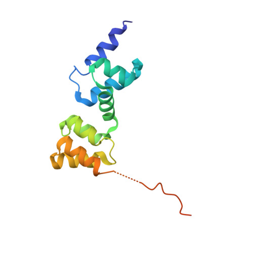
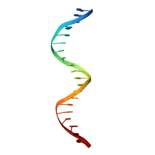
Diversification of DNA-Binding Specificity by Permissive and Specificity-Switching Mutations in the ParB/Noc Protein Family.

Jalal, A.S.B.Tran, N.T.Stevenson, C.E.Chan, E.W.Lo, R.Tan, X.Noy, A.Lawson, D.M.Le, T.B.K.

(2020) Cell Rep 32: 107928-107928

  • DOI: https://doi.org/10.1016/j.celrep.2020.107928
  • Primary Citation of Related Structures:  
    6S6H, 6Y93

  • PubMed Abstract: 

    Specific interactions between proteins and DNA are essential to many biological processes. Yet, it remains unclear how the diversification in DNA-binding specificity was brought about, and the mutational paths that led to changes in specificity are unknown. Using a pair of evolutionarily related DNA-binding proteins, each with a different DNA preference (ParB [Partitioning Protein B] and Noc [Nucleoid Occlusion Factor], which both play roles in bacterial chromosome maintenance), we show that specificity is encoded by a set of four residues at the protein-DNA interface. Combining X-ray crystallography and deep mutational scanning of the interface, we suggest that permissive mutations must be introduced before specificity-switching mutations to reprogram specificity and that mutational paths to new specificity do not necessarily involve dual-specificity intermediates. Overall, our results provide insight into the possible evolutionary history of ParB and Noc and, in a broader context, might be useful for understanding the evolution of other classes of DNA-binding proteins.


  • Organizational Affiliation

    Department of Molecular Microbiology, John Innes Centre, Norwich NR4 7UH, UK.


Macromolecules

Find similar proteins by:  (by identity cutoff)  |  3D Structure
Entity ID: 1
MoleculeChains Sequence LengthOrganismDetailsImage
Chromosome-partitioning protein ParB
A, B
143Caulobacter vibrioides NA1000Mutation(s): 0 
Gene Names: parBCCNA_03868
UniProt
Find proteins for B8GW30 (Caulobacter vibrioides (strain NA1000 / CB15N))
Explore B8GW30 
Go to UniProtKB:  B8GW30
Entity Groups  
Sequence Clusters30% Identity50% Identity70% Identity90% Identity95% Identity100% Identity
UniProt GroupB8GW30
Sequence Annotations
Expand
  • Reference Sequence

Find similar nucleic acids by:  Sequence   |   3D Structure  

Entity ID: 2
MoleculeChains LengthOrganismImage
DNA (5'-D(*GP*AP*TP*GP*TP*TP*TP*CP*AP*CP*GP*TP*GP*AP*AP*AP*CP*AP*TP*C)-3')
C, D
20Caulobacter vibrioides
Sequence Annotations
Expand
  • Reference Sequence
Experimental Data & Validation

Experimental Data

  • Method: X-RAY DIFFRACTION
  • Resolution: 2.40 Å
  • R-Value Free: 0.232 
  • R-Value Work: 0.216 
  • R-Value Observed: 0.217 
  • Space Group: C 1 2 1
Unit Cell:
Length ( Å )Angle ( ˚ )
a = 127.099α = 90
b = 40.717β = 121.4
c = 94.004γ = 90
Software Package:
Software NamePurpose
REFMACrefinement
DIALSdata scaling
PDB_EXTRACTdata extraction
DIALSdata reduction
PHASERphasing

Structure Validation

View Full Validation Report


Entry History & Funding Information

Deposition Data


Funding OrganizationLocationGrant Number
Royal SocietyUnited KingdomUF140053
Royal SocietyUnited KingdomRG150448
Biotechnology and Biological Sciences Research CouncilUnited KingdomBB/P018165/1
Biotechnology and Biological Sciences Research CouncilUnited KingdomBBS/E/J/000C0683
Biotechnology and Biological Sciences Research CouncilUnited KingdomBB/P012523/1

Revision History  (Full details and data files)

  • Version 1.0: 2020-07-15
    Type: Initial release
  • Version 1.1: 2021-01-27
    Changes: Database references
  • Version 1.2: 2024-01-24
    Changes: Data collection, Database references, Refinement description