6Y1Z | pdb_00006y1z

Mouse serotonin 5HT3 receptor in complex with palonosetron


Experimental Data Snapshot

  • Method: ELECTRON MICROSCOPY
  • Resolution: 2.82 Å
  • Aggregation State: PARTICLE 
  • Reconstruction Method: SINGLE PARTICLE 

wwPDB Validation 3D Report Full Report

Validation slider image for 6Y1Z

This is version 3.1 of the entry. See complete history

Literature

The Binding of Palonosetron and Other Antiemetic Drugs to the Serotonin 5-HT3 Receptor.

Zarkadas, E.Zhang, H.Cai, W.Effantin, G.Perot, J.Neyton, J.Chipot, C.Schoehn, G.Dehez, F.Nury, H.

(2020) Structure 28: 1131-1140.e4

  • DOI: https://doi.org/10.1016/j.str.2020.07.004
  • Primary Citation Related Structures: 
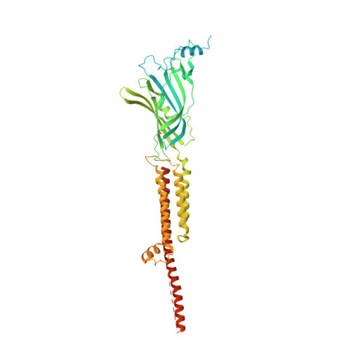
    6Y1Z

  • PubMed Abstract: 

    Inaccurately perceived as niche drugs, antiemetics are key elements of cancer treatment alleviating the most dreaded side effect of chemotherapy. Serotonin 5-HT3 receptor antagonists are the most commonly prescribed class of drugs to control chemotherapy-induced nausea and vomiting. These antagonists have been clinically successful drugs since the 1980s, yet our understanding of how they operate at the molecular level has been hampered by the difficulty of obtaining structures of drug-receptor complexes. Here, we report the cryoelectron microscopy structure of the palonosetron-bound 5-HT3 receptor. We investigate the binding of palonosetron, granisetron, dolasetron, ondansetron, and cilansetron using molecular dynamics, covering the whole set of antagonists used in clinical practice. The structural and computational results yield detailed atomic insight into the binding modes of the drugs. In light of our data, we establish a comprehensive framework underlying the inhibition mechanism by the -setron drug family.


  • Organizational Affiliation
    • CNRS, Univ. Grenoble Alpes, CEA, IBS, 38000 Grenoble, France.

Macromolecule Content 

  • Total Structure Weight: 327.86 kDa 
  • Atom Count: 16,610 
  • Modeled Residue Count: 1,920 
  • Deposited Residue Count: 2,830 
  • Unique protein chains: 1

Macromolecules

Find similar proteins by:|  3D Structure
Entity ID: 1
MoleculeChains  Sequence LengthOrganismDetailsImage
5-hydroxytryptamine receptor 3AA,
B [auth D],
C [auth B],
D [auth C],
E
566Mus musculusMutation(s): 0 
Gene Names: Htr3a5ht3Htr3
Membrane Entity: Yes 
UniProt & NIH Common Fund Data Resources
Find proteins for P23979 (Mus musculus)
Explore P23979 
Go to UniProtKB:  P23979
IMPC:  MGI:96282
Entity Groups
Sequence Clusters30% Identity50% Identity70% Identity90% Identity95% Identity100% Identity
UniProt GroupP23979
Glycosylation
Glycosylation Sites: 3Go to GlyGen: P23979-1
Sequence Annotations
Expand
Reference Sequence

Oligosaccharides

Help  
Entity ID: 2
MoleculeChains Length2D Diagram GlycosylationD Interactions
beta-D-mannopyranose-(1-4)-2-acetamido-2-deoxy-beta-D-glucopyranose-(1-4)-2-acetamido-2-deoxy-beta-D-glucopyranose
F, H, J, L, N
3N-Glycosylation
Glycosylation Resources
GlyTouCan: G15407YE
GlyCosmos: G15407YE
GlyGen: G15407YE
Entity ID: 3
MoleculeChains Length2D Diagram GlycosylationD Interactions
2-acetamido-2-deoxy-beta-D-glucopyranose-(1-4)-2-acetamido-2-deoxy-beta-D-glucopyranose
G, I, K, M, O
2N-Glycosylation
Glycosylation Resources
GlyTouCan: G42666HT
GlyCosmos: G42666HT
GlyGen: G42666HT

Small Molecules

Ligands 4 Unique
IDChains Name / Formula / InChI Key2D Diagram3D Interactions
O7B
(Subject of Investigation/LOI)

Query on O7B


Download:Ideal Coordinates CCD File
AA [auth B],
BA [auth C],
FA [auth E],
S [auth A],
W [auth D]
(3~{a}~{S})-2-[(3~{S})-1-azabicyclo[2.2.2]octan-3-yl]-3~{a},4,5,6-tetrahydro-3~{H}-benzo[de]isoquinolin-1-one
C19 H24 N2 O
CPZBLNMUGSZIPR-NVXWUHKLSA-N
NAG

Query on NAG


Download:Ideal Coordinates CCD File
EA [auth C],
IA [auth E],
R [auth A],
V [auth D],
Z [auth B]
2-acetamido-2-deoxy-beta-D-glucopyranose
C8 H15 N O6
OVRNDRQMDRJTHS-FMDGEEDCSA-N
TRP

Query on TRP


Download:Ideal Coordinates CCD File
CA [auth C],
GA [auth E],
P [auth A],
T [auth D],
X [auth B]
TRYPTOPHAN
C11 H12 N2 O2
QIVBCDIJIAJPQS-VIFPVBQESA-N
HIS

Query on HIS


Download:Ideal Coordinates CCD File
DA [auth C],
HA [auth E],
Q [auth A],
U [auth D],
Y [auth B]
HISTIDINE
C6 H10 N3 O2
HNDVDQJCIGZPNO-YFKPBYRVSA-O
Binding Affinity Annotations 
IDSourceBinding Affinity
O7B BindingDB:  6Y1Z Ki: 0.04 (nM) from 1 assay(s)

Experimental Data & Validation

Experimental Data

  • Method: ELECTRON MICROSCOPY
  • Resolution: 2.82 Å
  • Aggregation State: PARTICLE 
  • Reconstruction Method: SINGLE PARTICLE 
EM Software:
TaskSoftware PackageVersion
RECONSTRUCTIONcryoSPARCv2.13
MODEL REFINEMENTPHENIX1.17

Structure Validation

View Full Validation Report


Entry History 

& Funding Information

Deposition Data


Funding OrganizationLocationGrant Number
European Research Council (ERC)France637733

Revision History  (Full details and data files)

  • Version 1.0: 2020-03-04
    Type: Initial release
  • Version 2.0: 2020-07-29
    Type: Remediation
    Reason: Carbohydrate remediation
    Changes: Advisory, Atomic model, Data collection, Derived calculations, Structure summary
  • Version 2.1: 2020-08-12
    Changes: Database references, Structure summary
  • Version 2.2: 2020-10-14
    Changes: Database references
  • Version 3.0: 2022-09-14
    Changes: Advisory, Atomic model, Data collection, Database references, Derived calculations, Polymer sequence, Source and taxonomy, Structure summary
  • Version 3.1: 2024-11-06
    Changes: Data collection, Structure summary