6XI2

Apo form of POMGNT2


Experimental Data Snapshot

  • Method: X-RAY DIFFRACTION
  • Resolution: 2.57 Å
  • R-Value Free: 0.237 
  • R-Value Work: 0.213 
  • R-Value Observed: 0.214 

wwPDB Validation   3D Report Full Report


Ligand Structure Quality Assessment 


This is version 1.0 of the entry. See complete history


Literature

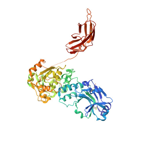
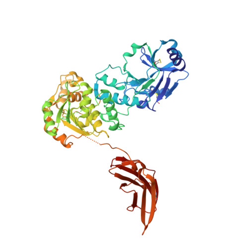
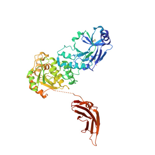
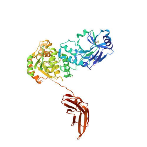
Crystal structures of beta-1,4-N-acetylglucosaminyltransferase 2: structural basis for inherited muscular dystrophies.

Yang, J.Y.Halmo, S.M.Praissman, J.Chapla, D.Singh, D.Wells, L.Moremen, K.W.Lanzilotta, W.N.

(2021) Acta Crystallogr D Struct Biol 77: 486-495

  • DOI: https://doi.org/10.1107/S2059798321001261
  • Primary Citation of Related Structures:  
    6XFI, 6XI2

  • PubMed Abstract: 

    The canonical O-mannosylation pathway in humans is essential for the functional glycosylation of α-dystroglycan. Disruption of this post-translational modification pathway leads to congenital muscular dystrophies. The first committed step in the construction of a functional matriglycan structure involves the post-translational modification of α-dystroglycan. This is essential for binding extracellular matrix proteins and arenaviruses, and is catalyzed by β-1,4-N-acetylglucosaminyltransferase 2 (POMGNT2). While another glycosyl transferase, β-1,4-N-acetylglucosaminyltransferase 1 (POMGNT1), has been shown to be promiscuous in extending O-mannosylated sites, POMGNT2 has been shown to display significant primary amino-acid selectivity near the site of O-mannosylation. Moreover, several single point mutations in POMGNT2 have been identified in patients with assorted dystroglycanopathies such as Walker-Warburg syndrome and limb girdle muscular dystrophy. To gain insight into POMGNT2 function in humans, the enzyme was expressed as a soluble, secreted fusion protein by transient infection of HEK293 suspension cultures. Here, crystal structures of POMGNT2 (amino-acid residues 25-580) with and without UDP bound are reported. Consistent with a novel fold and a unique domain organization, no molecular-replacement model was available and phases were obtained through crystallization of a selenomethionine variant of the enzyme in the same space group. Tetragonal (space group P42 1 2; unit-cell parameters a = b = 129.8, c = 81.6 Å, α = γ = β = 90°) crystals with UDP bound diffracted to 1.98 Å resolution and contained a single monomer in the asymmetric unit. Orthorhombic (space group P2 1 2 1 2 1 ; unit-cell parameters a = 142.3, b = 153.9, c = 187.4 Å, α = γ = β = 90°) crystals were also obtained; they diffracted to 2.57 Å resolution and contained four monomers with differential glycosylation patterns and conformations. These structures provide the first rational basis for an explanation of the loss-of-function mutations and offer significant insights into the mechanics of this important human enzyme.


  • Organizational Affiliation

    The Complex Carbohydrate Research Center, University of Georgia, Athens, GA 30602, USA.


Macromolecules
Find similar proteins by:  (by identity cutoff)  |  3D Structure
Entity ID: 1
MoleculeChains Sequence LengthOrganismDetailsImage
Protein O-linked-mannose beta-1,4-N-acetylglucosaminyltransferase 2533Homo sapiensMutation(s): 0 
Gene Names: POMGNT2AGO61C3orf39EOGTLGTDC2
EC: 2.4.1.312
UniProt & NIH Common Fund Data Resources
Find proteins for Q8NAT1 (Homo sapiens)
Explore Q8NAT1 
Go to UniProtKB:  Q8NAT1
PHAROS:  Q8NAT1
GTEx:  ENSG00000144647 
Entity Groups  
Sequence Clusters30% Identity50% Identity70% Identity90% Identity95% Identity100% Identity
UniProt GroupQ8NAT1
Sequence Annotations
Expand
  • Reference Sequence
Find similar proteins by:  (by identity cutoff)  |  3D Structure
Entity ID: 2
MoleculeChains Sequence LengthOrganismDetailsImage
Protein O-linked-mannose beta-1,4-N-acetylglucosaminyltransferase 2528Homo sapiensMutation(s): 0 
Gene Names: POMGNT2AGO61C3orf39EOGTLGTDC2
EC: 2.4.1.312
UniProt & NIH Common Fund Data Resources
Find proteins for Q8NAT1 (Homo sapiens)
Explore Q8NAT1 
Go to UniProtKB:  Q8NAT1
PHAROS:  Q8NAT1
GTEx:  ENSG00000144647 
Entity Groups  
Sequence Clusters30% Identity50% Identity70% Identity90% Identity95% Identity100% Identity
UniProt GroupQ8NAT1
Sequence Annotations
Expand
  • Reference Sequence
Find similar proteins by:  (by identity cutoff)  |  3D Structure
Entity ID: 3
MoleculeChains Sequence LengthOrganismDetailsImage
Protein O-linked-mannose beta-1,4-N-acetylglucosaminyltransferase 2C [auth D]533Homo sapiensMutation(s): 0 
Gene Names: POMGNT2AGO61C3orf39EOGTLGTDC2
EC: 2.4.1.312
UniProt & NIH Common Fund Data Resources
Find proteins for Q8NAT1 (Homo sapiens)
Explore Q8NAT1 
Go to UniProtKB:  Q8NAT1
PHAROS:  Q8NAT1
GTEx:  ENSG00000144647 
Entity Groups  
Sequence Clusters30% Identity50% Identity70% Identity90% Identity95% Identity100% Identity
UniProt GroupQ8NAT1
Sequence Annotations
Expand
  • Reference Sequence
Find similar proteins by:  (by identity cutoff)  |  3D Structure
Entity ID: 4
MoleculeChains Sequence LengthOrganismDetailsImage
Protein O-linked-mannose beta-1,4-N-acetylglucosaminyltransferase 2D [auth C]528Homo sapiensMutation(s): 0 
Gene Names: POMGNT2AGO61C3orf39EOGTLGTDC2
EC: 2.4.1.312
UniProt & NIH Common Fund Data Resources
Find proteins for Q8NAT1 (Homo sapiens)
Explore Q8NAT1 
Go to UniProtKB:  Q8NAT1
PHAROS:  Q8NAT1
GTEx:  ENSG00000144647 
Entity Groups  
Sequence Clusters30% Identity50% Identity70% Identity90% Identity95% Identity100% Identity
UniProt GroupQ8NAT1
Sequence Annotations
Expand
  • Reference Sequence

Find similar proteins by:  Sequence   |   3D Structure  

Entity ID: 5
MoleculeChains Sequence LengthOrganismDetailsImage
ALA-ALA-ALA-ALA-ALA-ALA-ALA-ALA-ALA-ALAE [auth G]10Homo sapiensMutation(s): 0 
Entity Groups  
Sequence Clusters30% Identity50% Identity70% Identity90% Identity95% Identity100% Identity
Sequence Annotations
Expand
  • Reference Sequence

Find similar proteins by:  Sequence   |   3D Structure  

Entity ID: 6
MoleculeChains Sequence LengthOrganismDetailsImage
ALA-GLY-ALA-GLY-ALA-ALA-ALA-ALA-ALA-ALAF [auth H]10Homo sapiensMutation(s): 0 
Entity Groups  
Sequence Clusters30% Identity50% Identity70% Identity90% Identity95% Identity100% Identity
Sequence Annotations
Expand
  • Reference Sequence
Small Molecules
Ligands 2 Unique
IDChains Name / Formula / InChI Key2D Diagram3D Interactions
NAG
Query on NAG

Download Ideal Coordinates CCD File 
G [auth A],
P [auth B],
W [auth D]
2-acetamido-2-deoxy-beta-D-glucopyranose
C8 H15 N O6
OVRNDRQMDRJTHS-FMDGEEDCSA-N
PO4
Query on PO4

Download Ideal Coordinates CCD File 
AA [auth D]
BA [auth D]
CA [auth D]
DA [auth C]
EA [auth C]
AA [auth D],
BA [auth D],
CA [auth D],
DA [auth C],
EA [auth C],
FA [auth C],
GA [auth C],
H [auth A],
HA [auth C],
I [auth A],
J [auth A],
K [auth A],
L [auth A],
M [auth A],
N [auth A],
O [auth A],
Q [auth B],
R [auth B],
S [auth B],
T [auth B],
U [auth B],
V [auth B],
X [auth D],
Y [auth D],
Z [auth D]
PHOSPHATE ION
O4 P
NBIIXXVUZAFLBC-UHFFFAOYSA-K
Experimental Data & Validation

Experimental Data

  • Method: X-RAY DIFFRACTION
  • Resolution: 2.57 Å
  • R-Value Free: 0.237 
  • R-Value Work: 0.213 
  • R-Value Observed: 0.214 
  • Space Group: P 21 21 21
Unit Cell:
Length ( Å )Angle ( ˚ )
a = 142.322α = 90
b = 153.979β = 90
c = 187.494γ = 90
Software Package:
Software NamePurpose
PHENIXrefinement
PDB_EXTRACTdata extraction
HKL-2000data reduction
HKL-2000data scaling
PHASERphasing

Structure Validation

View Full Validation Report


Ligand Structure Quality Assessment 


Entry History & Funding Information

Deposition Data


Funding OrganizationLocationGrant Number
National Institutes of Health/National Institute of General Medical Sciences (NIH/NIGMS)United States--

Revision History  (Full details and data files)

  • Version 1.0: 2021-04-21
    Type: Initial release