6XGY

Crystal structure of E. coli MlaFB ABC transport subunits in the dimeric state


Experimental Data Snapshot

  • Method: X-RAY DIFFRACTION
  • Resolution: 2.90 Å
  • R-Value Free: 0.243 
  • R-Value Work: 0.196 
  • R-Value Observed: 0.198 

wwPDB Validation   3D Report Full Report


Ligand Structure Quality Assessment 


This is version 1.1 of the entry. See complete history


Literature

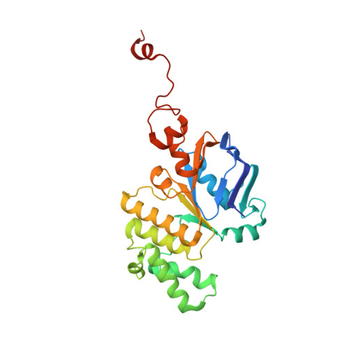
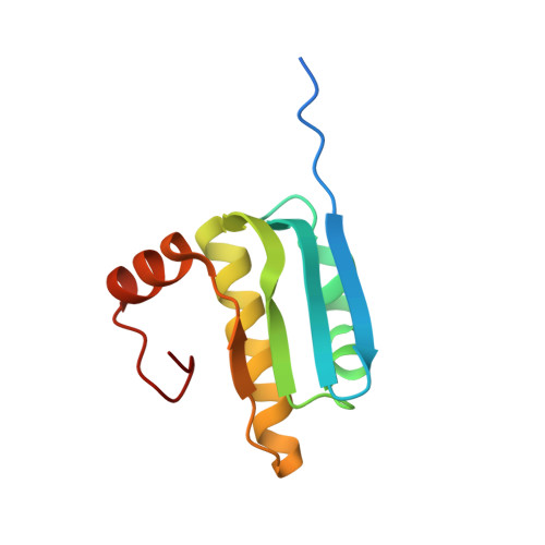
Structure of MlaFB uncovers novel mechanisms of ABC transporter regulation.

Kolich, L.R.Chang, Y.T.Coudray, N.Giacometti, S.I.MacRae, M.R.Isom, G.L.Teran, E.M.Bhabha, G.Ekiert, D.C.

(2020) Elife 9

  • DOI: https://doi.org/10.7554/eLife.60030
  • Primary Citation of Related Structures:  
    6XGY, 6XGZ

  • PubMed Abstract: 

    ABC transporters facilitate the movement of diverse molecules across cellular membranes, but how their activity is regulated post-translationally is not well understood. Here we report the crystal structure of MlaFB from E. coli , the cytoplasmic portion of the larger MlaFEDB ABC transporter complex, which drives phospholipid trafficking across the bacterial envelope to maintain outer membrane integrity. MlaB, a STAS domain protein, binds the ABC nucleotide binding domain, MlaF, and is required for its stability. Our structure also implicates a unique C-terminal tail of MlaF in self-dimerization. Both the C-terminal tail of MlaF and the interaction with MlaB are required for the proper assembly of the MlaFEDB complex and its function in cells. This work leads to a new model for how an important bacterial lipid transporter may be regulated by small proteins, and raises the possibility that similar regulatory mechanisms may exist more broadly across the ABC transporter family.


  • Organizational Affiliation

    Department of Cell Biology, New York University School of Medicine, New York, United States.


Macromolecules
Find similar proteins by:  (by identity cutoff)  |  3D Structure
Entity ID: 1
MoleculeChains Sequence LengthOrganismDetailsImage
Organic solvent ABC transporter ATP-binding protein269Escherichia coli LAU-EC10Mutation(s): 0 
Gene Names: V415_03760
Entity Groups  
Sequence Clusters30% Identity50% Identity70% Identity90% Identity95% Identity100% Identity
Sequence Annotations
Expand
  • Reference Sequence
Find similar proteins by:  (by identity cutoff)  |  3D Structure
Entity ID: 2
MoleculeChains Sequence LengthOrganismDetailsImage
ABC transporter maintaining OM lipid asymmetry, cytoplasmic STAS component109Escherichia coliMutation(s): 0 
Gene Names: mlaByrbB
UniProt
Find proteins for P64602 (Escherichia coli (strain K12))
Explore P64602 
Go to UniProtKB:  P64602
Entity Groups  
Sequence Clusters30% Identity50% Identity70% Identity90% Identity95% Identity100% Identity
UniProt GroupP64602
Sequence Annotations
Expand
  • Reference Sequence
Experimental Data & Validation

Experimental Data

  • Method: X-RAY DIFFRACTION
  • Resolution: 2.90 Å
  • R-Value Free: 0.243 
  • R-Value Work: 0.196 
  • R-Value Observed: 0.198 
  • Space Group: P 32 2 1
Unit Cell:
Length ( Å )Angle ( ˚ )
a = 102.71α = 90
b = 102.71β = 90
c = 89.71γ = 120
Software Package:
Software NamePurpose
PHENIXrefinement
PDB_EXTRACTdata extraction
XDSdata reduction
XSCALEdata scaling
PHASERphasing

Structure Validation

View Full Validation Report


Ligand Structure Quality Assessment 


Entry History & Funding Information

Deposition Data


Funding OrganizationLocationGrant Number
National Institutes of Health/National Institute of General Medical Sciences (NIH/NIGMS)United StatesR35GM128777
National Institutes of Health/National Institute of General Medical Sciences (NIH/NIGMS)United StatesR00GM112982
Other privateUnited StatesDFS-20-16
American Heart AssociationUnited States20POST35210202
National Institutes of Health/National Institute of General Medical Sciences (NIH/NIGMS)United StatesT32 GM088118

Revision History  (Full details and data files)

  • Version 1.0: 2020-07-15
    Type: Initial release
  • Version 1.1: 2023-10-18
    Changes: Data collection, Database references, Refinement description